Pfam Domains mapped on to the structure: 2ILK
No.
Chain ID
Pfam ID
Pfam Description
Linkout - Pfam
Linkout - CDD
1
A
PF00726
Interleukin 10
PF00726
PF00726
Conserved Domain Database Superfamily Annotations: 2ILK
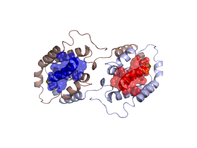
Structural Details of PDB entry 2ILK
Structural Details of PDB entry 2ILK
PDBid Chains Hinge Swapped Domain
2ILK
A,B
A:141-144,B:141-144
A:145-160,B:145-160
Swapped-domain interface residues and interactions:
Chains Residues
A
53 , 56 , 57 , 145 , 146 , 147 , 148 , 149 , 150 , 151 , 152 , 153 , 154 , 156 , 157 , 158 , 160 ,
B
53 , 56 , 57 , 145 , 146 , 147 , 148 , 149 , 150 , 151 , 152 , 153 , 154 , 156 , 157 , 158 ,
Non-swapped-domain interface residues and interactions:
Chains Residues
A
6 , 7 , 16 , 19 , 20 , 23 , 26 , 27 , 30 , 37 , 38 , 40 , 41 , 42 , 43 , 45 , 46 , 47 , 48 , 51 , 52 , 55 , 59 , 60 , 61 , 62 , 63 , 64 , 65 , 67 , 68 , 71 , 72 , 75 , 76 , 77 , 79 , 80 , 83 , 94 , 98 , 101 , 105 , 110 , 111 , 113 , 114 , 116 , 117 , 118 , 120 , 121 , 123 , 124 , 125 , 127 , 128 , 131 , 134 , 136 , 137 , 138 , 139 , 140 , 141 , 142 , 143 , 144 ,
B
6 , 7 , 16 , 19 , 20 , 23 , 26 , 27 , 30 , 37 , 38 , 40 , 41 , 42 , 43 , 45 , 46 , 47 , 48 , 51 , 52 , 55 , 59 , 60 , 61 , 62 , 63 , 64 , 65 , 67 , 68 , 71 , 72 , 75 , 76 , 77 , 79 , 80 , 83 , 94 , 98 , 101 , 105 , 110 , 111 , 113 , 114 , 116 , 117 , 118 , 120 , 121 , 123 , 124 , 125 , 127 , 128 , 131 , 134 , 136 , 137 , 138 , 139 , 140 , 141 , 142 , 143 , 144 ,
Mutations in critical regions:
Chains
Hinge
Domain swapped interface Non-swapped interface Swapped Domain
A No mutation No mutation No mutation No mutation B No mutation No mutation No mutation No mutation
HIDE output:
JMOL Visualization:
2D-plot:
JOY Structural annotation for hinge hinge and swapped domain:
JOY output: