Home BrowseSearch Help Group

Pfam Domains mapped on to the structure: 2I9F

No. Chain ID Pfam ID Pfam Description Linkout - Pfam Linkout - CDD
1 A PF01481 Arterivirus nucleocapsid protein PF01481 PF01481

Gene Ontology Annotations: 2I9F

No. GO ID GO Description Linkout - AmiGO
1 GO:0019013 viral nucleocapsid GO:0019013

Conserved Domain Database Superfamily Annotations: 2I9F

No. PDB ID PSSM ID CDD Accession Superfamily Short Name Linkout - CDD
1 2I9F 110480 Arteri_nucleo superfamily N -
Structural Details of PDB entry 2I9F

Structural Details of PDB entry 2I9F

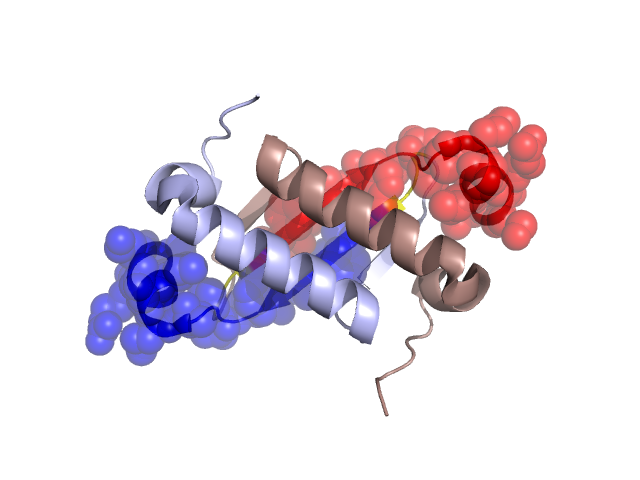
PDBid Chains Hinge Swapped Domain
2I9F A,B A:86-88,B:86-88 A:89-105,B:89-105

Swapped-domain interface residues and interactions:

Chains Residues
A 83, 86, 87, 88, 89, 90, 91, 92, 93, 94, 95, 96, 104, 105,
B 87, 88, 89, 90, 91, 92, 93, 94, 95, 96, 104,

Non-swapped-domain interface residues and interactions:

Chains Residues
A 7, 8, 51, 53, 54, 55, 63, 66, 67, 68, 70, 71, 72, 74, 76, 77, 79, 81, 82,
B 7, 8, 51, 53, 54, 55, 63, 66, 67, 68, 70, 71, 72, 74, 76, 77, 79, 80, 81, 82, 83, 86,

Swapped domains are represented using trasperent spheres.
Non-swapped part is reprented using light color and cartoon reprentation.
Hinge region is shown in yellow color.

Mutations in critical regions:

Chains Hinge Domain swapped interface Non-swapped interface Swapped Domain
ANo mutationNo mutationNo mutationNo mutation
BNo mutationNo mutationNo mutationNo mutation

HIDE output:

Homologues found through HIDE algorithm

JMOL Visualization:

Jmol_logo Jmol_logo

2D-plot:

3:2I9F A:2I9F B:2I9F

JOY Structural annotation for hinge hinge and swapped domain:

Hinge Swapped domain

JOY output:

ali file:2I9F.ali
atm file:2I9F.atm
cof file:2I9F.cof
hbd file:2I9F.hbd
html file:2I9F.html
pdb file:2I9F.pdb
ps file:2I9F.ps
psa file:2I9F.psa
rtf file:2I9F.rtf
sst file:2I9F.sst
tem file:2I9F.tem