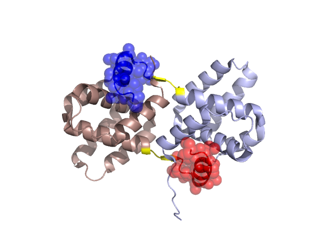
Pfam Domains mapped on to the structure: 2I8B
No.
Chain ID
Pfam ID
Pfam Description
Linkout - Pfam
Linkout - CDD
1
A
PF11507
Ebola virus-specific transcription factor VP30
PF11507
PF11507
Conserved Domain Database Superfamily Annotations: 2I8B
Structural Details of PDB entry 2I8B
Structural Details of PDB entry 2I8B
PDBid Chains Hinge Swapped Domain
2I8B
A,B
A:248-251,B:248-251
A:252-266,B:252-266
Swapped-domain interface residues and interactions:
Chains Residues
A
140 , 141 , 142 , 150 , 169 , 173 , 252 , 253 , 255 , 257 , 258 , 260 , 261 , 262 , 263 , 264 , 265 , 266 ,
B
141 , 142 , 150 , 153 , 169 , 173 , 252 , 253 , 257 , 258 , 260 , 261 , 262 , 263 , 264 , 265 ,
Non-swapped-domain interface residues and interactions:
Chains Residues
A
143 , 144 , 145 , 146 , 147 , 148 , 149 , 152 , 153 , 154 , 166 , 172 , 176 , 179 , 180 , 181 , 185 , 246 , 247 , 248 , 249 , 250 , 251 ,
B
139 , 140 , 143 , 144 , 145 , 146 , 147 , 148 , 149 , 152 , 154 , 166 , 172 , 176 , 180 , 181 , 185 , 246 , 247 , 248 , 249 , 250 , 251 ,
Mutations in critical regions:
Chains
Hinge
Domain swapped interface Non-swapped interface Swapped Domain
A No mutation GLY(140)-, ALA(141)-, No mutation No mutation B No mutation ALA(141)-, GLN(139)-, GLY(140)-, No mutation
HIDE output:
JMOL Visualization:
2D-plot:
JOY Structural annotation for hinge hinge and swapped domain:
JOY output: