Pfam Domains mapped on to the structure: 2HZL
No.
Chain ID
Pfam ID
Pfam Description
Linkout - Pfam
Linkout - CDD
1
A
PF03480
Bacterial extracellular solute-binding protein, family 7
PF03480
PF03480
Gene Ontology Annotations: 2HZL
No.
GO ID
GO Description
Linkout - AmiGO
1
GO:0030288
outer membrane-bounded periplasmic space
GO:0030288
Conserved Domain Database Superfamily Annotations: 2HZL
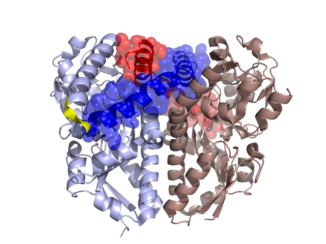
Structural Details of PDB entry 2HZL
Structural Details of PDB entry 2HZL
PDBid Chains Hinge Swapped Domain
2HZL
A,B
A:323-325,B:323-325
A:326-365,B:326-365
Swapped-domain interface residues and interactions:
Chains Residues
A
155 , 157 , 235 , 236 , 241 , 285 , 337 , 338 , 339 , 340 , 341 , 342 , 343 , 344 , 345 , 346 , 347 , 348 , 349 , 350 , 351 , 352 , 353 , 354 , 355 , 356 , 357 , 359 , 360 , 361 , 364 , 365 ,
B
155 , 157 , 235 , 236 , 241 , 285 , 288 , 337 , 338 , 339 , 340 , 341 , 342 , 343 , 344 , 345 , 346 , 347 , 348 , 349 , 350 , 351 , 352 , 353 , 354 , 355 , 356 , 357 , 359 , 360 , 361 , 364 ,
Non-swapped-domain interface residues and interactions:
Chains Residues
A
57 , 60 , 61 , 63 , 116 , 117 , 119 , 120 , 121 , 124 , 125 , 129 , 130 , 154 , 218 , 260 , 263 , 264 , 267 , 268 , 270 , 271 , 274 , 275 , 277 , 278 , 279 , 281 , 282 , 284 , 286 , 288 , 289 , 298 , 306 , 307 , 310 ,
B
57 , 60 , 61 , 63 , 116 , 117 , 119 , 120 , 121 , 124 , 125 , 129 , 130 , 154 , 218 , 260 , 263 , 264 , 267 , 268 , 270 , 271 , 274 , 275 , 277 , 278 , 279 , 281 , 282 , 284 , 286 , 289 , 298 , 300 , 306 , 307 , 310 ,
Mutations in critical regions:
Chains
Hinge
Domain swapped interface Non-swapped interface Swapped Domain
A No mutation No mutation No mutation No mutation B No mutation No mutation No mutation No mutation
HIDE output:
JMOL Visualization:
2D-plot:
JOY Structural annotation for hinge hinge and swapped domain:
JOY output: