Home BrowseSearch Help Group

Pfam Domains mapped on to the structure: 2HT0

No. Chain ID Pfam ID Pfam Description Linkout - Pfam Linkout - CDD
1 A PF00216 Bacterial DNA-binding protein PF00216 PF00216
2 B PF00216 Bacterial DNA-binding protein PF00216 PF00216

Conserved Domain Database Superfamily Annotations: 2HT0

No. PDB ID PSSM ID CDD Accession Superfamily Short Name Linkout - CDD
1 2HT0 29683 cd00591 HU_IHF - cl00257
2 2HT0 206937 cl00257 HU_IHF superfamily - -
Structural Details of PDB entry 2HT0

Structural Details of PDB entry 2HT0

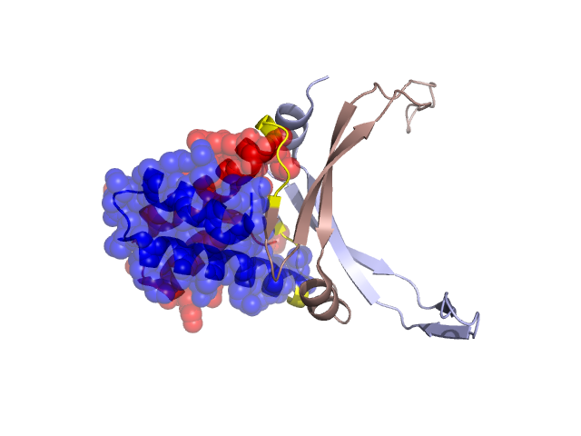
PDBid Chains Hinge Swapped Domain
2HT0 A,B A:39-44,B:38-43 A:2-38,B:1-37

Swapped-domain interface residues and interactions:

Chains Residues
A 2, 3, 4, 5, 7, 8, 11, 12, 15, 16, 18, 22, 25, 26, 29, 30, 31, 32, 33, 34, 35, 36, 37, 38, 42, 43, 44, 45, 46, 47, 87, 91,
B 1, 2, 3, 6, 9, 10, 13, 14, 16, 17, 21, 22, 24, 25, 28, 29, 30, 31, 32, 33, 34, 36, 37, 41, 43, 44, 45, 48, 86, 90,

Non-swapped-domain interface residues and interactions:

Chains Residues
A 39, 49, 52, 56, 75, 76, 77, 79, 81, 83, 88, 90, 92, 94, 95, 96, 97,
B 38, 39, 40, 42, 47, 51, 53, 75, 76, 77, 78, 80, 82, 85, 89, 91, 92,

Swapped domains are represented using trasperent spheres.
Non-swapped part is represented using light color and cartoon representation.
Hinge region is shown in yellow color.

Mutations in critical regions:

Chains Hinge Domain swapped interface Non-swapped interface Swapped Domain
ANo mutationNo mutationNo mutationNo mutation
BNo mutationNo mutationNo mutationNo mutation

HIDE output:

Homologues found through HIDE algorithm

JMOL Visualization:

Jmol_logo            Jmol_logo            Jmol_logo

2D-plot:

A:2HT0 B:2HT0

JOY Structural annotation for hinge hinge and swapped domain:

Hinge Swapped domain

JOY output:

ali file:2HT0.ali
atm file:2HT0.atm
cof file:2HT0.cof
hbd file:2HT0.hbd
html file:2HT0.html
pdb file:2HT0.pdb
ps file:2HT0.ps
psa file:2HT0.psa
rtf file:2HT0.rtf
sst file:2HT0.sst
tem file:2HT0.tem