Pfam Domains mapped on to the structure: 2HS1
No.
Chain ID
Pfam ID
Pfam Description
Linkout - Pfam
Linkout - CDD
1
A
PF00077
Retroviral aspartyl protease
PF00077
PF00077
Conserved Domain Database Superfamily Annotations: 2HS1
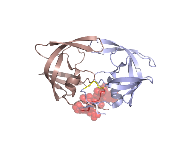
Structural Details of PDB entry 2HS1
Structural Details of PDB entry 2HS1
PDBid Chains Hinge Swapped Domain
2HS1
A,B
A:7-9,B:7-9;A:107-109,B:107-109
A:1-6,B:1-6
Swapped-domain interface residues and interactions:
Chains Residues
A
1 , 2 , 3 , 4 , 5 , 6 ,
B
187 , 191 , 195 , 196 , 197 ,
Non-swapped-domain interface residues and interactions:
Chains Residues
A
8 , 9 , 23 , 24 , 25 , 26 , 27 , 29 , 48 , 49 , 50 , 51 , 52 , 54 , 67 , 69 , 81 , 84 , 87 , 90 , 91 , 93 , 94 , 95 , 96 , 97 , 98 , 99 ,
B
101 , 102 , 103 , 104 , 105 , 106 , 107 , 108 , 109 , 123 , 124 , 125 , 126 , 127 , 129 , 147 , 149 , 150 , 151 , 152 , 154 , 167 , 169 , 181 , 184 , 190 , 192 , 193 , 194 , 198 ,
Mutations in critical regions:
Chains
Hinge
Domain swapped interface Non-swapped interface Swapped Domain
A LYS(7)GLN, No mutation ALA(67)CYS, ALA(95)CYS, No mutation B LYS(107)GLN, ALA(195)CYS, LYS(107)GLN, ALA(167)CYS, No mutation B LYS(107)GLN, ALA(195)CYS, LYS(107)GLN, ALA(167)CYS, No mutation
HIDE output:
JMOL Visualization:
2D-plot:
JOY Structural annotation for hinge hinge and swapped domain:
JOY output: