Pfam Domains mapped on to the structure: 2HN1
No.
Chain ID
Pfam ID
Pfam Description
Linkout - Pfam
Linkout - CDD
1
A
PF01544
CorA-like Mg2+ transporter protein
PF01544
PF01544
Gene Ontology Annotations: 2HN1
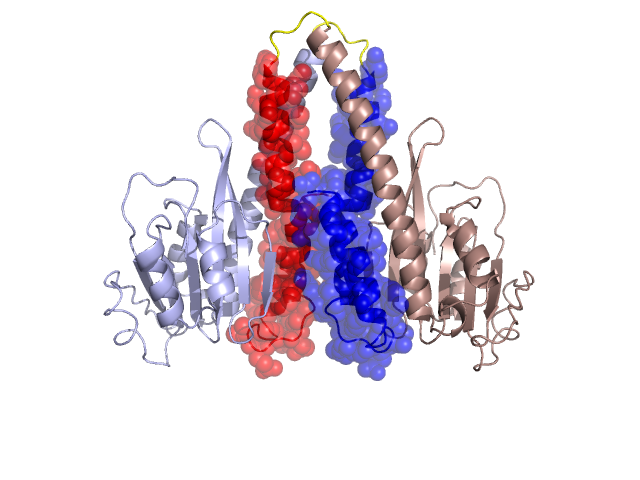
Structural Details of PDB entry 2HN1
Structural Details of PDB entry 2HN1
PDBid Chains Hinge Swapped Domain
2HN1
A,B
A:203-208,B:203-208
A:209-263,B:209-263
Swapped-domain interface residues and interactions:
Chains Residues
A
102 , 104 , 116 , 124 , 135 , 168 , 169 , 172 , 176 , 179 , 183 , 210 , 211 , 214 , 217 , 218 , 220 , 221 , 222 , 223 , 224 , 225 , 226 , 227 , 228 , 229 , 230 , 231 , 232 , 233 , 234 , 235 , 237 , 238 , 240 , 242 , 243 , 244 , 246 , 247 , 250 , 251 , 253 , 254 , 257 , 258 , 261 , 262 , 263 ,
B
102 , 104 , 116 , 124 , 135 , 168 , 169 , 172 , 176 , 179 , 183 , 210 , 211 , 214 , 217 , 218 , 220 , 221 , 222 , 223 , 224 , 225 , 226 , 227 , 228 , 229 , 230 , 231 , 232 , 233 , 234 , 235 , 237 , 238 , 240 , 242 , 243 , 244 , 246 , 247 , 250 , 251 , 253 , 254 , 257 , 258 , 261 , 262 ,
Non-swapped-domain interface residues and interactions:
Chains Residues
A
101 , 106 , 108 , 111 , 113 , 115 , 118 , 126 , 133 , 165 , 171 , 175 , 180 , 182 , 186 , 189 , 192 , 193 , 196 , 199 , 200 , 208 ,
B
101 , 106 , 108 , 111 , 113 , 115 , 118 , 126 , 133 , 165 , 171 , 175 , 180 , 182 , 186 , 189 , 192 , 193 , 196 , 199 , 200 , 208 ,
Mutations in critical regions:
Chains
Hinge
Domain swapped interface Non-swapped interface Swapped Domain
A No mutation No mutation No mutation No mutation B No mutation No mutation No mutation No mutation
HIDE output:
JMOL Visualization:
2D-plot:
JOY Structural annotation for hinge hinge and swapped domain:
JOY output: