Home BrowseSearch Help Group

Pfam Domains mapped on to the structure: 2HMW

No. Chain ID Pfam ID Pfam Description Linkout - Pfam Linkout - CDD
1 A PF03446 NAD binding domain of 6-phosphogluconate dehydrogenase PF03446 PF03446
2 A PF02254 TrkA-N domain PF02254 PF02254
3 A PF13460 NADH(P)-binding PF13460 PF13460

Conserved Domain Database Superfamily Annotations: 2HMW

No. PDB ID PSSM ID CDD Accession Superfamily Short Name Linkout - CDD
1 2HMW 212616 cl09931 NADB_Rossmann superfamily - -
Structural Details of PDB entry 2HMW

Structural Details of PDB entry 2HMW

PDBid Chains Hinge Swapped Domain
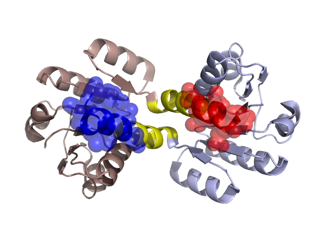
2HMW A,B A:124-133,B:124-133 A:134-140,B:134-144

Swapped-domain interface residues and interactions:

Chains Residues
A 134, 135, 136, 137, 139, 140,
B 134, 135, 136, 137, 139, 140, 141,

Non-swapped-domain interface residues and interactions:

Chains Residues
A 8, 9, 16, 17, 20, 21, 24, 25, 28, 30, 73, 75, 101, 103, 105, 120, 122, 124, 125, 126, 127, 128, 129, 130, 131, 132, 133,
B 8, 9, 16, 17, 20, 21, 24, 25, 28, 30, 73, 75, 101, 103, 105, 106, 120, 122, 124, 125, 126, 128, 129, 130, 131, 132, 133,

Swapped domains are represented using trasperent spheres.
Non-swapped part is represented using light color and cartoon representation.
Hinge region is shown in yellow color.

Mutations in critical regions:

Chains Hinge Domain swapped interface Non-swapped interface Swapped Domain
ANo mutationNo mutationNo mutationNo mutation
BNo mutationNo mutationNo mutationNo mutation

HIDE output:

Homologues found through HIDE algorithm

JMOL Visualization:

Jmol_logo            Jmol_logo            Jmol_logo

2D-plot:

A:2HMW B:2HMW

JOY Structural annotation for hinge hinge and swapped domain:

Hinge Swapped domain

JOY output:

ali file:2HMW.ali
atm file:2HMW.atm
cof file:2HMW.cof
hbd file:2HMW.hbd
html file:2HMW.html
pdb file:2HMW.pdb
ps file:2HMW.ps
psa file:2HMW.psa
rtf file:2HMW.rtf
sst file:2HMW.sst
tem file:2HMW.tem