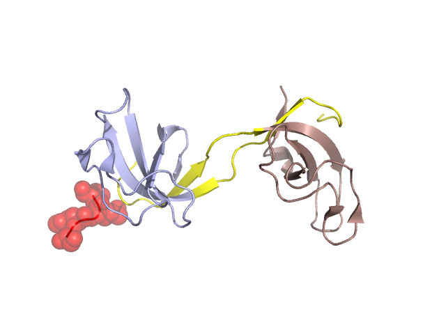
Pfam Domains mapped on to the structure: 2HL3 No. Chain ID Pfam ID Pfam Description Linkout - Pfam Linkout - CDD 1 A PF01302 CAP-Gly domain PF01302 PF01302 Structural Details of PDB entry 2HL3 Structural Details of PDB entry 2HL3 PDBid Chains Hinge Swapped Domain 2HL3 A,B A:26-40,B:26-40 A:16-25,B:26-25 Swapped-domain interface residues and interactions: Chains Residues A 17, 18, 19, B 45, 78, 79, 80, Non-swapped-domain interface residues and interactions: Chains Residues A 27, 28, 29, 30, 31, 32, 33, 34, 35, 36, 37, 38, 39, 40, 41, 42, 43, 44, 45, 47, 60, 62, 63, 68, 87, 91, 92, 93, 94, 95, 96, 97, B 27, 29, 30, 31, 32, 33, 34, 35, 36, 37, 38, 39, 40, 41, 42, 43, 44, 47, 60, 61, 62, 63, 87, 89, 91, 93, 94, 95, 96, Swapped domains are represented using trasperent spheres. Non-swapped part is represented using light color and cartoon representation. Hinge region is shown in yellow color. Mutations in critical regions: Chains Hinge Domain swapped interface Non-swapped interface Swapped Domain ANo mutationHIS(17)-, No mutationSER(16)-, HIS(17)-, BNo mutationNo mutationNo mutationNo mutation HIDE output: Homologues found through HIDE algorithm JMOL Visualization: 2D-plot: A:2HL3 B:2HL3 JOY Structural annotation for hinge hinge and swapped domain: Hinge Swapped domain JOY output: ali file:2HL3.ali atm file:2HL3.atm cof file:2HL3.cof hbd file:2HL3.hbd html file:2HL3.html pdb file:2HL3.pdb ps file:2HL3.ps psa file:2HL3.psa rtf file:2HL3.rtf sst file:2HL3.sst tem file:2HL3.tem