Structural Details of PDB entry 2HH0
Structural Details of PDB entry 2HH0
PDBid Chains Hinge Swapped Domain
2HH0
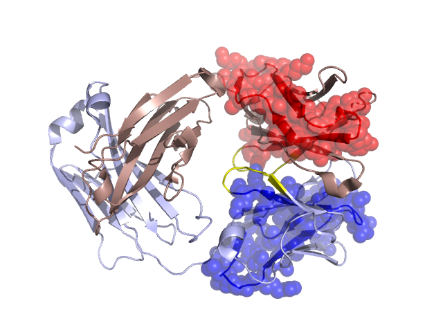
H,L
H:48-53,L:48-53
H:1-47,L:2-47
Swapped-domain interface residues and interactions:
Non-swapped-domain interface residues and interactions:
Chains Residues
H
51 , 52 , 54 , 69 , 71 , 72 , 105 , 134 , 135 , 137 , 139 , 141 , 160 , 161 , 162 , 163 , 165 , 166 , 167 , 168 , 178 , 182 , 184 , 211 , 212 , 213 , 214 , 216 , 217 , 218 , 232 , 234 , 236 , 267 ,
L
50 , 51 , 52 , 54 , 57 , 58 , 70 , 71 , 72 , 105 , 107 , 135 , 136 , 137 , 139 , 141 , 158 , 160 , 161 , 162 , 163 , 165 , 167 , 168 , 174 , 176 , 178 , 180 , 182 , 184 , 185 , 211 , 212 , 213 , 214 , 215 , 218 , 230 , 232 , 234 , 236 , 267 , 271 ,