Pfam Domains mapped on to the structure: 2H4P
Conserved Domain Database Superfamily Annotations: 2H4P
No.
PDB ID
PSSM ID
CDD Accession
Superfamily Short Name
Linkout - CDD
1
2H4P
206855
SERPIN
superfamily
N -
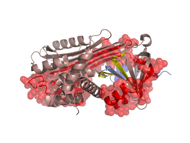
Structural Details of PDB entry 2H4P
Structural Details of PDB entry 2H4P
PDBid Chains Hinge Swapped Domain
2H4P
A,B
A:252-260,B:397-405
A:261-369,B:406-409
Swapped-domain interface residues and interactions:
Chains Residues
A
26 , 27 , 215 , 221 , 223 , 261 , 263 , 264 , 269 , 271 , 274 , 275 , 278 , 280 , 283 , 287 , 288 , 295 , 296 , 297 , 298 , 299 , 300 , 301 , 302 , 304 , 306 , 360 , 369 ,
B
378 , 379 , 380 , 381 , 382 , 383 , 386 , 406 , 407 , 408 ,
Non-swapped-domain interface residues and interactions:
Chains Residues
A
5 , 8 , 9 , 12 , 16 , 25 , 28 , 29 , 30 , 31 , 32 , 33 , 101 , 104 , 105 , 114 , 193 , 195 , 212 , 213 , 214 , 225 , 229 , 234 , 236 , 243 , 245 , 252 , 253 , 254 , 255 , 256 , 257 , 258 , 259 , 260 ,
B
377 , 384 , 385 , 387 , 388 , 389 , 390 , 391 , 392 , 393 , 394 , 396 , 397 , 398 , 399 , 400 , 401 , 402 , 403 , 404 , 405 ,
Mutations in critical regions:
Chains
Hinge
Domain swapped interface Non-swapped interface Swapped Domain
A No mutation No mutation No mutation No mutation B No mutation No mutation No mutation No mutation
HIDE output:
JMOL Visualization:
2D-plot:
JOY Structural annotation for hinge hinge and swapped domain:
JOY output: