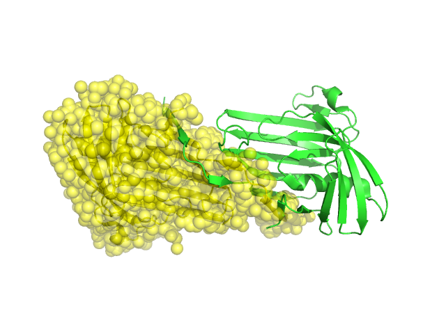
Structural Details of PDB entry 2H1T
Structural Details of PDB entry 2H1T
PDBid Chains Hinge Swapped Domain
2H1T
A,B
A:-,B:-
A:2--1,B:4--1
Swapped-domain interface residues and interactions:
Non-swapped-domain interface residues and interactions:
Chains Residues
A
3 , 4 , 5 , 6 , 7 , 8 , 9 , 10 , 11 , 12 , 13 , 14 , 15 , 16 , 17 , 18 , 19 , 20 , 21 , 22 , 23 , 24 , 25 , 26 , 35 , 36 , 37 , 39 , 42 , 43 , 44 , 46 , 67 , 68 , 69 , 105 , 108 , 109 , 176 , 177 , 182 , 183 , 184 , 185 , 186 , 187 ,
B
4 , 5 , 6 , 7 , 8 , 9 , 10 , 11 , 12 , 13 , 14 , 15 , 16 , 17 , 18 , 19 , 20 , 21 , 22 , 23 , 24 , 25 , 26 , 35 , 36 , 37 , 39 , 42 , 43 , 44 , 46 , 67 , 68 , 69 , 105 , 108 , 109 , 172 , 174 , 176 , 177 , 182 , 183 , 184 , 185 , 186 ,