Home BrowseSearch Help Group

Pfam Domains mapped on to the structure: 2GUD

No. Chain ID Pfam ID Pfam Description Linkout - Pfam Linkout - CDD
1 A PF01419 Jacalin-like lectin domain PF01419 PF01419

Conserved Domain Database Superfamily Annotations: 2GUD

No. PDB ID PSSM ID CDD Accession Superfamily Short Name Linkout - CDD
1 2GUD 187710 cd09614 griffithsin_like - cl03205
2 2GUD 198810 cl03205 Jacalin_like superfamily - -
Structural Details of PDB entry 2GUD

Structural Details of PDB entry 2GUD

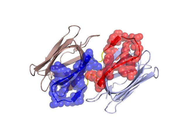
PDBid Chains Hinge Swapped Domain
2GUD A,B A:16-19,B:16-19 A:1-15,B:1-15

Swapped-domain interface residues and interactions:

Chains Residues
A 1, 2, 3, 4, 5, 6, 7, 8, 9, 11, 12, 13, 14, 15, 65, 66, 67, 69, 104, 105, 106, 107, 108, 112, 113, 114, 115, 116, 117, 118, 119, 120, 121,
B 1, 2, 3, 4, 5, 6, 7, 8, 9, 11, 12, 13, 14, 15, 65, 66, 67, 69, 104, 105, 106, 107, 108, 112, 113, 114, 115, 116, 117, 118, 119, 120,

Non-swapped-domain interface residues and interactions:

Chains Residues
A 16, 17, 19, 34, 35, 37, 39, 63, 93, 94, 95, 101, 102,
B 16, 17, 19, 34, 35, 37, 39, 63, 93, 95, 99, 101, 102,

Swapped domains are represented using trasperent spheres.
Non-swapped part is represented using light color and cartoon representation.
Hinge region is shown in yellow color.

Mutations in critical regions:

Chains Hinge Domain swapped interface Non-swapped interface Swapped Domain
ANo mutationNo mutationNo mutationNo mutation
BNo mutationNo mutationNo mutationNo mutation

HIDE output:

Homologues found through HIDE algorithm

JMOL Visualization:

Jmol_logo            Jmol_logo            Jmol_logo

2D-plot:

A:2GUD B:2GUD

JOY Structural annotation for hinge hinge and swapped domain:

Hinge Swapped domain

JOY output:

ali file:2GUD.ali
atm file:2GUD.atm
cof file:2GUD.cof
hbd file:2GUD.hbd
html file:2GUD.html
pdb file:2GUD.pdb
ps file:2GUD.ps
psa file:2GUD.psa
rtf file:2GUD.rtf
sst file:2GUD.sst
tem file:2GUD.tem