Home BrowseSearch Help Group

Pfam Domains mapped on to the structure: 2GBO

No. Chain ID Pfam ID Pfam Description Linkout - Pfam Linkout - CDD
1 A PF07408 Protein of unknown function (DUF1507) PF07408 PF07408

Conserved Domain Database Superfamily Annotations: 2GBO

No. PDB ID PSSM ID CDD Accession Superfamily Short Name Linkout - CDD
1 2GBO 187070 cl11487 DUF1507 superfamily - -
Structural Details of PDB entry 2GBO

Structural Details of PDB entry 2GBO

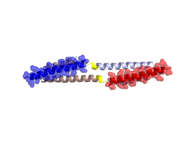
PDBid Chains Hinge Swapped Domain
2GBO A,B A:31-34,B:31-34 A:2-30,B:2-30

Swapped-domain interface residues and interactions:

Chains Residues
A 7, 10, 11, 14, 15, 17, 18, 21, 22, 24, 25, 26, 27, 28, 29, 41, 44, 63, 76, 79, 80,
B 7, 10, 11, 14, 15, 17, 18, 21, 22, 24, 25, 26, 27, 28, 29, 41, 44, 63, 76, 79, 80,

Non-swapped-domain interface residues and interactions:

Chains Residues
A 33, 34, 37, 38, 40, 47, 51, 54, 58, 62, 64, 72, 75, 82,
B 33, 34, 37, 38, 40, 47, 51, 54, 58, 62, 64, 72, 75,

Swapped domains are represented using trasperent spheres.
Non-swapped part is represented using light color and cartoon representation.
Hinge region is shown in yellow color.

Mutations in critical regions:

Chains Hinge Domain swapped interface Non-swapped interface Swapped Domain
ANo mutationNo mutationNo mutationNo mutation
BNo mutationNo mutationNo mutationNo mutation

HIDE output:

Homologues found through HIDE algorithm

JMOL Visualization:

Jmol_logo            Jmol_logo            Jmol_logo

2D-plot:

A:2GBO B:2GBO

JOY Structural annotation for hinge hinge and swapped domain:

Hinge Swapped domain

JOY output:

ali file:2GBO.ali
atm file:2GBO.atm
cof file:2GBO.cof
hbd file:2GBO.hbd
html file:2GBO.html
pdb file:2GBO.pdb
ps file:2GBO.ps
psa file:2GBO.psa
rtf file:2GBO.rtf
sst file:2GBO.sst
tem file:2GBO.tem