Pfam Domains mapped on to the structure: 2FUR
No.
Chain ID
Pfam ID
Pfam Description
Linkout - Pfam
Linkout - CDD
1
A
PF12900
Pyridoxamine 5'-phosphate oxidase
PF12900
PF12900
Conserved Domain Database Superfamily Annotations: 2FUR
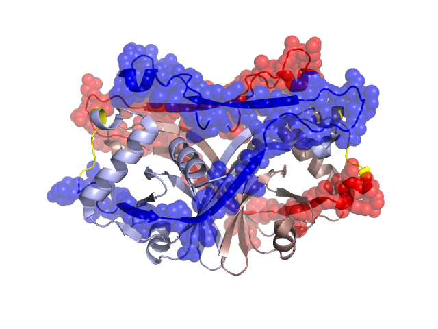
Structural Details of PDB entry 2FUR
Structural Details of PDB entry 2FUR
PDBid Chains Hinge Swapped Domain
2FUR
A,B
A:137-142,B:137-142
A:143-208,B:143-208
Swapped-domain interface residues and interactions:
Chains Residues
A
137 , 138 , 145 , 172 , 174 , 176 , 177 , 180 , 181 , 182 , 183 , 184 , 185 , 186 , 187 , 188 , 189 , 190 , 193 , 194 , 195 , 198 , 206 , 207 , 208 ,
B
138 , 145 , 174 , 176 , 177 , 180 , 181 , 182 , 183 , 184 , 185 , 186 , 187 , 188 , 189 , 190 , 193 , 194 , 195 , 198 , 206 , 207 ,
Non-swapped-domain interface residues and interactions:
Chains Residues
A
32 , 33 , 35 , 37 , 39 , 42 , 43 , 44 , 45 , 46 , 48 , 77 , 79 , 81 , 83 , 85 , 88 , 90 , 91 , 92 , 93 , 94 , 95 , 97 , 99 , 103 , 105 , 107 , 123 , 127 , 130 , 131 , 134 , 139 , 141 ,
B
32 , 33 , 35 , 37 , 39 , 42 , 43 , 44 , 45 , 46 , 48 , 77 , 79 , 81 , 83 , 85 , 88 , 90 , 91 , 92 , 93 , 94 , 95 , 97 , 99 , 103 , 105 , 107 , 123 , 127 , 130 , 131 , 134 , 137 , 139 , 141 ,
Mutations in critical regions:
Chains
Hinge
Domain swapped interface Non-swapped interface Swapped Domain
A No mutation No mutation No mutation No mutation B No mutation No mutation No mutation No mutation
HIDE output:
JMOL Visualization:
2D-plot:
JOY Structural annotation for hinge hinge and swapped domain:
JOY output: