Home BrowseSearch Help Group

Pfam Domains mapped on to the structure: 2FPN

No. Chain ID Pfam ID Pfam Description Linkout - Pfam Linkout - CDD
1 A PF08680 Protein of unknown function (DUF1779) PF08680 PF08680
Structural Details of PDB entry 2FPN

Structural Details of PDB entry 2FPN

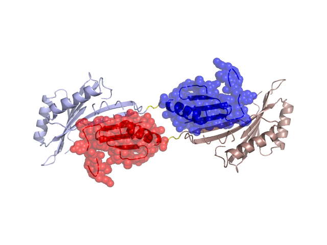
PDBid Chains Hinge Swapped Domain
2FPN A,B A:136-139,B:136-139 A:140-214,B:140-214

Swapped-domain interface residues and interactions:

Chains Residues
A 3, 10, 14, 128, 129, 130, 131, 132, 133, 134, 135, 146, 149, 150, 153, 154, 177, 179, 203, 205, 206, 207, 208, 209, 210, 211, 212, 213, 214,
B 3, 10, 14, 128, 129, 130, 131, 132, 133, 134, 135, 146, 149, 150, 153, 154, 177, 179, 203, 205, 206, 207, 208, 209, 210, 211, 212, 213,

Non-swapped-domain interface residues and interactions:

Chains Residues
A 6, 13, 15, 16, 24, 126, 127,
B 6, 13, 15, 16, 24, 126, 127,

Swapped domains are represented using trasperent spheres.
Non-swapped part is represented using light color and cartoon representation.
Hinge region is shown in yellow color.

Mutations in critical regions:

Chains Hinge Domain swapped interface Non-swapped interface Swapped Domain
ANo mutationNo mutationNo mutationNo mutation
BNo mutationNo mutationNo mutationNo mutation

HIDE output:

Homologues found through HIDE algorithm

JMOL Visualization:

Jmol_logo            Jmol_logo            Jmol_logo

2D-plot:

A:2FPN B:2FPN

JOY Structural annotation for hinge hinge and swapped domain:

Hinge Swapped domain

JOY output:

ali file:2FPN.ali
atm file:2FPN.atm
cof file:2FPN.cof
hbd file:2FPN.hbd
html file:2FPN.html
pdb file:2FPN.pdb
ps file:2FPN.ps
psa file:2FPN.psa
rtf file:2FPN.rtf
sst file:2FPN.sst
tem file:2FPN.tem