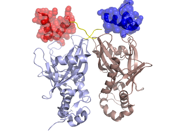
Structural Details of PDB entry 2FML
Structural Details of PDB entry 2FML
PDBid Chains Hinge Swapped Domain
2FML
A,B
A:33-38,B:33-38
A:3-32,B:4-32
Swapped-domain interface residues and interactions:
Chains Residues
A
15 , 16 , 18 , 20 , 23 , 24 , 27 , 28 , 64 , 65 , 66 , 67 , 120 ,
B
15 , 16 , 18 , 20 , 23 , 24 , 27 , 28 , 64 , 65 , 66 , 67 , 120 ,
Non-swapped-domain interface residues and interactions:
Chains Residues
A
35 , 36 , 37 , 38 , 39 , 40 , 69 , 78 , 79 , 80 , 81 , 82 , 83 , 84 , 85 , 86 , 108 , 110 , 111 , 113 , 115 , 119 , 121 , 122 , 124 , 126 , 141 , 142 , 200 , 203 , 268 ,
B
35 , 36 , 37 , 38 , 39 , 40 , 69 , 78 , 79 , 81 , 82 , 83 , 84 , 85 , 86 , 108 , 110 , 111 , 113 , 115 , 119 , 121 , 122 , 124 , 126 , 196 , 203 ,