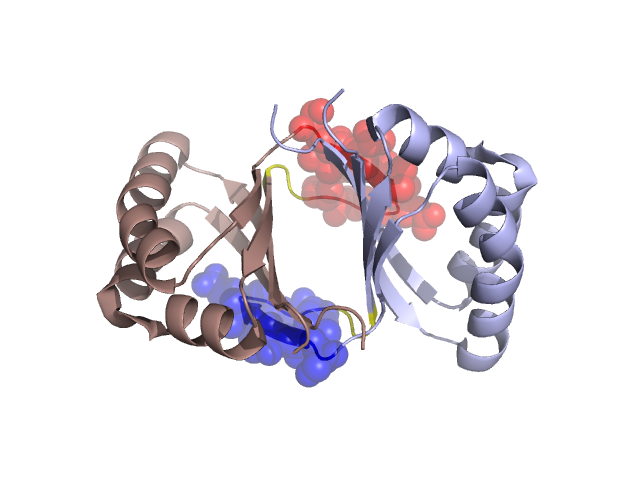
Structural Details of PDB entry 2FIU
Structural Details of PDB entry 2FIU
PDBid Chains Hinge Swapped Domain
2FIU
A,B
A:39-41,B:39-41;A:51-53,B:51-53
A:42-50,B:42-50
Swapped-domain interface residues and interactions:
Chains Residues
A
42 , 43 , 44 , 46 , 47 , 48 , 49 , 88 , 91 , 92 , 93 ,
B
42 , 43 , 44 , 46 , 47 , 48 , 49 , 88 , 91 , 92 , 93 ,
Non-swapped-domain interface residues and interactions:
Chains Residues
A
5 , 7 , 9 , 36 , 37 , 38 , 39 , 41 , 54 , 56 , 62 , 66 , 69 , 81 , 82 , 87 , 94 , 95 , 96 ,
B
5 , 7 , 9 , 36 , 37 , 38 , 39 , 41 , 54 , 56 , 62 , 66 , 69 , 81 , 82 , 87 , 94 , 95 ,