Home BrowseSearch Help Group

Pfam Domains mapped on to the structure: 2F1R

No. Chain ID Pfam ID Pfam Description Linkout - Pfam Linkout - CDD
1 A PF03205 Molybdopterin guanine dinucleotide synthesis protein B PF03205 PF03205
2 A PF01656 CobQ/CobB/MinD/ParA nucleotide binding domain PF01656 PF01656

Conserved Domain Database Superfamily Annotations: 2F1R

No. PDB ID PSSM ID CDD Accession Superfamily Short Name Linkout - CDD
1 2F1R 48380 cd03116 MobB - cl09099
2 2F1R 212613 cl09099 P-loop_NTPase superfamily - -
Structural Details of PDB entry 2F1R

Structural Details of PDB entry 2F1R

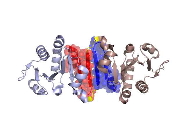
PDBid Chains Hinge Swapped Domain
2F1R A,B A:76-78,B:76-78 A:51-75,B:51-75

Swapped-domain interface residues and interactions:

Chains Residues
A 20, 23, 24, 27, 33, 34, 35, 36, 37, 38, 52, 53, 55, 56, 57, 59, 60, 61, 62, 63, 64, 65, 66, 67, 68, 69, 70, 71, 72, 73, 74, 75, 76, 77, 79, 82,
B 16, 20, 23, 24, 33, 34, 35, 36, 37, 38, 39, 40, 52, 53, 55, 56, 57, 59, 60, 61, 62, 63, 64, 65, 66, 67, 68, 69, 70, 71, 72, 73, 74, 75, 76, 77, 79, 82, 83,

Non-swapped-domain interface residues and interactions:

Chains Residues
A 16, 19, 32, 88, 91, 92, 93, 94, 95, 96, 162,
B 13, 19, 27, 88, 91, 92, 93, 95, 96,

Swapped domains are represented using trasperent spheres.
Non-swapped part is represented using light color and cartoon representation.
Hinge region is shown in yellow color.

Mutations in critical regions:

Chains Hinge Domain swapped interface Non-swapped interface Swapped Domain
ANo mutationNo mutationGLU(162)-, No mutation
BNo mutationNo mutationNo mutationNo mutation

HIDE output:

Homologues found through HIDE algorithm

JMOL Visualization:

Jmol_logo            Jmol_logo            Jmol_logo

2D-plot:

A:2F1R B:2F1R

JOY Structural annotation for hinge hinge and swapped domain:

Hinge Swapped domain

JOY output:

ali file:2F1R.ali
atm file:2F1R.atm
cof file:2F1R.cof
hbd file:2F1R.hbd
html file:2F1R.html
pdb file:2F1R.pdb
ps file:2F1R.ps
psa file:2F1R.psa
rtf file:2F1R.rtf
sst file:2F1R.sst
tem file:2F1R.tem