Pfam Domains mapped on to the structure: 2ES0
No.
Chain ID
Pfam ID
Pfam Description
Linkout - Pfam
Linkout - CDD
1
A
PF00615
Regulator of G protein signaling domain
PF00615
PF00615
Conserved Domain Database Superfamily Annotations: 2ES0
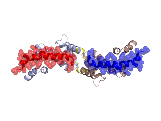
Structural Details of PDB entry 2ES0
Structural Details of PDB entry 2ES0
PDBid Chains Hinge Swapped Domain
2ES0
A,B
A:414-419,B:414-419
A:420-452,B:420-452
Swapped-domain interface residues and interactions:
Chains Residues
A
327 , 330 , 334 , 337 , 350 , 353 , 362 , 366 , 369 , 373 , 402 , 406 , 410 , 413 , 414 , 420 , 421 , 422 , 424 , 425 , 426 , 428 , 429 , 430 , 432 , 433 , 434 , 437 , 438 , 440 , 441 , 442 , 444 , 446 , 447 , 450 , 451 , 452 ,
B
327 , 330 , 334 , 337 , 350 , 353 , 362 , 366 , 369 , 373 , 402 , 406 , 410 , 413 , 414 , 420 , 421 , 422 , 424 , 425 , 426 , 428 , 429 , 430 , 432 , 433 , 434 , 437 , 438 , 440 , 441 , 442 , 444 , 446 , 447 , 450 , 451 ,
Non-swapped-domain interface residues and interactions:
Chains Residues
A
324 , 331 , 333 , 336 , 338 , 340 , 341 , 349 , 354 , 357 , 359 , 365 , 372 , 375 , 377 , 380 , 381 , 384 , 385 , 387 , 401 , 403 , 409 , 417 , 418 , 419 ,
B
324 , 331 , 333 , 336 , 338 , 340 , 341 , 349 , 354 , 357 , 359 , 365 , 372 , 375 , 377 , 380 , 381 , 384 , 385 , 387 , 401 , 403 , 409 , 417 , 418 , 419 ,
Mutations in critical regions:
Chains
Hinge
Domain swapped interface Non-swapped interface Swapped Domain
A No mutation No mutation No mutation No mutation B No mutation No mutation No mutation No mutation
HIDE output:
JMOL Visualization:
2D-plot:
JOY Structural annotation for hinge hinge and swapped domain:
JOY output: