Home BrowseSearch Help Group

Pfam Domains mapped on to the structure: 2EHJ

No. Chain ID Pfam ID Pfam Description Linkout - Pfam Linkout - CDD
1 A PF00156 Phosphoribosyl transferase domain PF00156 PF00156

Conserved Domain Database Superfamily Annotations: 2EHJ

No. PDB ID PSSM ID CDD Accession Superfamily Short Name Linkout - CDD
1 2EHJ 206754 cd06223 PRTases_typeI - cl00309
2 2EHJ 212186 cl00309 PRTases_typeI superfamily - -
Structural Details of PDB entry 2EHJ

Structural Details of PDB entry 2EHJ

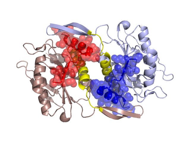
PDBid Chains Hinge Swapped Domain
2EHJ A,D A:25-49,D:25-49 A:1-24,D:1-24

Swapped-domain interface residues and interactions:

Chains Residues
A 8, 9, 10, 12, 13, 14, 16, 19, 20, 21, 45, 47, 49, 54, 55, 56, 57, 63, 64, 65, 66,
D 8, 9, 10, 12, 13, 14, 16, 19, 20, 21, 45, 47, 49, 54, 55, 56, 57, 63, 64, 65, 66,

Non-swapped-domain interface residues and interactions:

Chains Residues
A 27, 34, 38, 41, 42, 44, 58, 61, 89, 90, 91, 92, 188, 194, 195, 198, 203, 208,
D 27, 34, 38, 41, 42, 44, 58, 61, 89, 90, 92, 188, 192, 194, 195, 196, 197, 198,

Swapped domains are represented using trasperent spheres.
Non-swapped part is represented using light color and cartoon representation.
Hinge region is shown in yellow color.

Mutations in critical regions:

Chains Hinge Domain swapped interface Non-swapped interface Swapped Domain
ANo mutationNo mutationNo mutationNo mutation
DNo mutationNo mutationNo mutationNo mutation

HIDE output:

Homologues found through HIDE algorithm

JMOL Visualization:

Jmol_logo            Jmol_logo            Jmol_logo

2D-plot:

A:2EHJ D:2EHJ

JOY Structural annotation for hinge hinge and swapped domain:

Hinge Swapped domain

JOY output:

ali file:2EHJ.ali
atm file:2EHJ.atm
cof file:2EHJ.cof
hbd file:2EHJ.hbd
html file:2EHJ.html
pdb file:2EHJ.pdb
ps file:2EHJ.ps
psa file:2EHJ.psa
rtf file:2EHJ.rtf
sst file:2EHJ.sst
tem file:2EHJ.tem