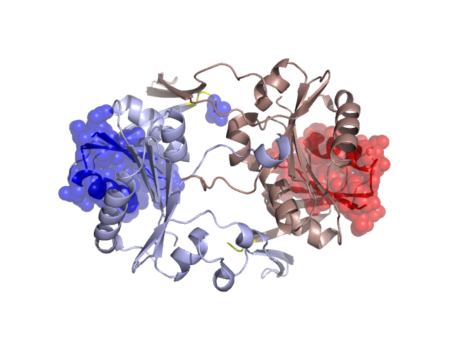
Pfam Domains mapped on to the structure: 2E6E
No.
Chain ID
Pfam ID
Pfam Description
Linkout - Pfam
Linkout - CDD
1
A
PF01975
Survival protein SurE
PF01975
PF01975
Conserved Domain Database Superfamily Annotations: 2E6E
Structural Details of PDB entry 2E6E
Structural Details of PDB entry 2E6E
PDBid Chains Hinge Swapped Domain
2E6E
A,B
A:47-49,B:47-49
A:1-46,B:1-46
Swapped-domain interface residues and interactions:
Non-swapped-domain interface residues and interactions:
Chains Residues
A
47 , 48 , 49 , 50 , 51 , 52 , 53 , 76 , 79 , 80 , 83 , 84 , 103 , 105 , 106 , 115 , 116 , 119 , 120 , 155 , 156 , 158 , 159 , 174 , 175 , 176 , 177 , 179 , 181 , 182 , 184 , 186 , 191 , 195 , 196 , 197 , 198 , 199 , 200 , 226 , 227 , 228 , 229 , 230 , 231 , 232 , 233 , 234 , 235 , 236 ,
B
48 , 49 , 50 , 51 , 52 , 53 , 79 , 80 , 83 , 84 , 115 , 116 , 119 , 120 , 121 , 155 , 156 , 158 , 159 , 176 , 177 , 179 , 182 , 183 , 184 , 186 , 191 , 196 , 197 , 198 , 199 , 200 , 201 , 202 , 226 , 227 , 228 , 229 , 230 , 231 , 232 , 233 , 234 , 236 , 237 ,
Mutations in critical regions:
Chains
Hinge
Domain swapped interface Non-swapped interface Swapped Domain
A No mutation No mutation No mutation No mutation B No mutation No mutation No mutation No mutation
HIDE output:
JMOL Visualization:
2D-plot:
JOY Structural annotation for hinge hinge and swapped domain:
JOY output: