Home BrowseSearch Help Group

Pfam Domains mapped on to the structure: 2E55

No. Chain ID Pfam ID Pfam Description Linkout - Pfam Linkout - CDD
1 A PF00156 Phosphoribosyl transferase domain PF00156 PF00156

Conserved Domain Database Superfamily Annotations: 2E55

No. PDB ID PSSM ID CDD Accession Superfamily Short Name Linkout - CDD
1 2E55 206754 cd06223 PRTases_typeI - cl00309
2 2E55 212186 cl00309 PRTases_typeI superfamily - -
Structural Details of PDB entry 2E55

Structural Details of PDB entry 2E55

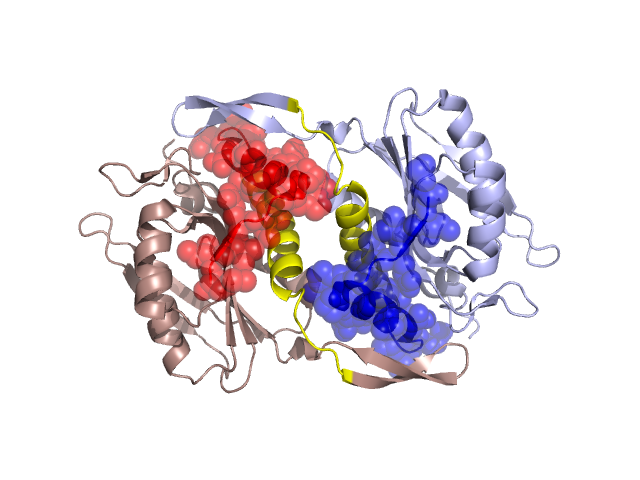
PDBid Chains Hinge Swapped Domain
2E55 A,D A:24-49,D:24-49 A:1-23,D:1-23

Swapped-domain interface residues and interactions:

Chains Residues
A 7, 8, 9, 11, 12, 13, 15, 18, 19, 20, 21, 44, 46, 48, 53, 54, 55, 56, 62, 63, 64, 65,
D 6, 7, 8, 9, 11, 12, 13, 15, 18, 19, 20, 21, 44, 46, 53, 54, 55, 56, 62, 63, 64, 65,

Non-swapped-domain interface residues and interactions:

Chains Residues
A 26, 29, 33, 36, 37, 40, 41, 43, 57, 60, 67, 88, 89, 91, 92, 190, 192, 193, 196, 208,
D 26, 29, 33, 36, 37, 40, 41, 43, 48, 57, 60, 67, 88, 89, 91, 92, 192, 193, 195, 196,

Swapped domains are represented using trasperent spheres.
Non-swapped part is represented using light color and cartoon representation.
Hinge region is shown in yellow color.

Mutations in critical regions:

Chains Hinge Domain swapped interface Non-swapped interface Swapped Domain
ANo mutationNo mutationNo mutationNo mutation
DNo mutationNo mutationNo mutationNo mutation

HIDE output:

Homologues found through HIDE algorithm

JMOL Visualization:

Jmol_logo            Jmol_logo            Jmol_logo

2D-plot:

A:2E55 D:2E55

JOY Structural annotation for hinge hinge and swapped domain:

Hinge Swapped domain

JOY output:

ali file:2E55.ali
atm file:2E55.atm
cof file:2E55.cof
hbd file:2E55.hbd
html file:2E55.html
pdb file:2E55.pdb
ps file:2E55.ps
psa file:2E55.psa
rtf file:2E55.rtf
sst file:2E55.sst
tem file:2E55.tem