Pfam Domains mapped on to the structure: 2D7V
No.
Chain ID
Pfam ID
Pfam Description
Linkout - Pfam
Linkout - CDD
1
A
PF02566
OsmC-like protein
PF02566
PF02566
Conserved Domain Database Superfamily Annotations: 2D7V
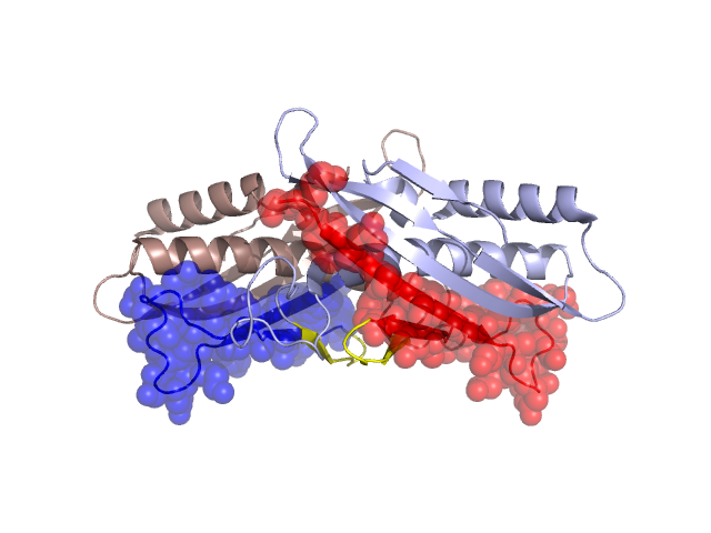
Structural Details of PDB entry 2D7V
Structural Details of PDB entry 2D7V
PDBid Chains Hinge Swapped Domain
2D7V
A,B
A:39-46,B:39-46
A:7-38,B:9-38
Swapped-domain interface residues and interactions:
Chains Residues
A
7 , 9 , 10 , 11 , 12 , 13 , 14 , 15 , 16 , 17 , 18 , 19 , 24 , 25 , 26 , 28 , 30 , 32 , 34 , 36 , 38 , 51 , 66 , 67 , 70 , 71 , 74 , 75 , 83 , 86 , 87 , 89 , 91 , 92 , 93 , 94 , 95 , 96 , 97 , 98 , 99 , 100 , 101 , 102 , 103 ,
B
9 , 10 , 11 , 12 , 13 , 14 , 15 , 16 , 17 , 18 , 19 , 24 , 25 , 26 , 28 , 30 , 32 , 34 , 36 , 38 , 51 , 66 , 67 , 70 , 71 , 74 , 75 , 83 , 86 , 89 , 91 , 92 , 93 , 94 , 95 , 96 , 97 , 98 , 99 , 100 , 101 , 102 , 103 ,
Non-swapped-domain interface residues and interactions:
Chains Residues
A
39 , 40 , 42 , 44 , 48 , 52 , 60 , 61 , 63 , 65 , 68 , 69 , 72 , 76 , 79 , 82 , 110 , 111 , 112 , 113 , 144 , 145 , 147 , 148 , 150 , 151 , 152 , 153 , 161 ,
B
40 , 42 , 44 , 48 , 49 , 52 , 60 , 61 , 63 , 65 , 68 , 69 , 72 , 76 , 79 , 82 , 110 , 111 , 112 , 113 , 144 , 147 , 148 , 150 , 151 , 152 , 153 ,
Mutations in critical regions:
Chains
Hinge
Domain swapped interface Non-swapped interface Swapped Domain
A No mutation No mutation No mutation No mutation B No mutation No mutation No mutation No mutation
HIDE output:
JMOL Visualization:
2D-plot:
JOY Structural annotation for hinge hinge and swapped domain:
JOY output: