Pfam Domains mapped on to the structure: 2D6Y
No.
Chain ID
Pfam ID
Pfam Description
Linkout - Pfam
Linkout - CDD
1
A
PF00440
Bacterial regulatory proteins, tetR family
PF00440
PF00440
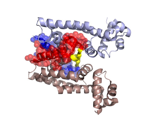
Structural Details of PDB entry 2D6Y
Structural Details of PDB entry 2D6Y
PDBid Chains Hinge Swapped Domain
2D6Y
A,B
A:157-161,B:157-161
A:162-191,B:162-191
Swapped-domain interface residues and interactions:
Chains Residues
A
103 , 107 , 108 , 118 , 121 , 141 , 144 , 145 , 162 , 164 , 165 , 166 , 167 , 168 , 177 , 178 , 181 , 182 , 184 , 185 , 188 , 189 , 191 ,
B
103 , 107 , 108 , 118 , 121 , 141 , 144 , 145 , 162 , 164 , 165 , 166 , 167 , 168 , 177 , 178 , 181 , 182 , 184 , 185 , 188 , 189 ,
Non-swapped-domain interface residues and interactions:
Chains Residues
A
99 , 100 , 101 , 102 , 109 , 111 , 114 , 117 , 125 , 138 , 140 , 142 , 147 , 148 , 149 , 151 , 152 , 155 , 156 , 157 , 159 , 160 , 161 , 192 ,
B
99 , 100 , 101 , 109 , 111 , 114 , 117 , 138 , 140 , 147 , 148 , 149 , 151 , 152 , 155 , 156 , 157 , 159 , 160 , 161 ,
Mutations in critical regions:
Chains
Hinge
Domain swapped interface Non-swapped interface Swapped Domain
A No mutation No mutation No mutation No mutation B No mutation No mutation No mutation No mutation
HIDE output:
JMOL Visualization:
2D-plot:
JOY Structural annotation for hinge hinge and swapped domain:
JOY output: