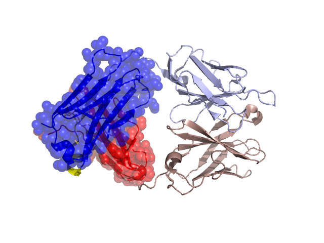
Structural Details of PDB entry 2D03
Structural Details of PDB entry 2D03
PDBid Chains Hinge Swapped Domain
2D03
H,L
H:131-140,L:125-134
H:141-220,L:135-218
Swapped-domain interface residues and interactions:
Chains Residues
H
143 , 145 , 147 , 149 , 170 , 171 , 172 , 173 , 175 , 177 , 184 , 186 , 214 , 219 ,
L
122 , 137 , 139 , 141 , 143 , 144 , 166 , 167 , 168 , 169 , 170 , 173 , 180 , 182 , 184 , 186 , 218 ,
Non-swapped-domain interface residues and interactions:
Chains Residues
H
36 , 40 , 45 , 46 , 48 , 59 , 61 , 62 , 95 , 100 , 102 , 103 , 104 , 105 , 106 , 107 , 109 , 110 , 128 , 129 , 130 , 131 , 132 , 133 ,
L
38 , 40 , 42 , 44 , 49 , 50 , 52 , 55 , 56 , 61 , 62 , 93 , 95 , 97 , 100 , 101 , 102 , 104 , 120 , 124 , 125 , 126 , 127 , 129 , 130 , 133 ,