Pfam Domains mapped on to the structure: 2CGR
No.
Chain ID
Pfam ID
Pfam Description
Linkout - Pfam
Linkout - CDD
1
H
PF13895
Immunoglobulin domain
PF13895
PF13895
Conserved Domain Database Superfamily Annotations: 2CGR
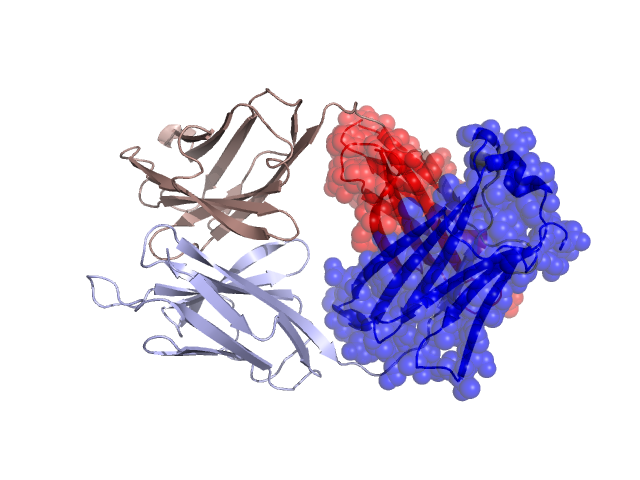
Structural Details of PDB entry 2CGR
Structural Details of PDB entry 2CGR
PDBid Chains Hinge Swapped Domain
2CGR
H,L
H:128-134,L:124-130
H:135-214,L:131-219
Swapped-domain interface residues and interactions:
Chains Residues
H
131 , 132 , 140 , 144 , 146 , 167 , 168 , 169 , 170 , 172 , 174 , 179 , 181 , 183 , 211 ,
L
132 , 136 , 138 , 140 , 142 , 143 , 165 , 166 , 167 , 168 , 169 , 179 , 181 , 183 , 185 , 214 , 218 , 219 ,
Non-swapped-domain interface residues and interactions:
Chains Residues
H
39 , 44 , 45 , 47 , 50 , 59 , 61 , 95 , 101 , 102 , 103 , 104 , 105 , 106 , 107 , 108 , 125 , 126 , 127 , 128 , 129 , 130 , 133 ,
L
1 , 37 , 39 , 41 , 43 , 48 , 49 , 50 , 51 , 60 , 92 , 96 , 99 , 100 , 101 , 102 , 103 , 105 , 119 , 121 , 122 , 123 , 124 , 126 , 128 , 129 ,
Mutations in critical regions:
Chains
Hinge
Domain swapped interface Non-swapped interface Swapped Domain
H No mutation No mutation GLU(39)GLN, ASN(59)LYS, ARG(61)ASN, SER(101)ALA, SER(102)PRO, No mutation L No mutation No mutation HIS(39)TYR, LEU(51)PRO, No mutation
HIDE output:
JMOL Visualization:
2D-plot:
JOY Structural annotation for hinge hinge and swapped domain:
JOY output: