Pfam Domains mapped on to the structure: 2C3B
No.
Chain ID
Pfam ID
Pfam Description
Linkout - Pfam
Linkout - CDD
1
A
PF00160
Cyclophilin type peptidyl-prolyl cis-trans isomerase/CLD
PF00160
PF00160
Conserved Domain Database Superfamily Annotations: 2C3B
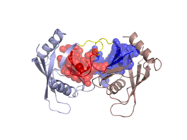
Structural Details of PDB entry 2C3B
Structural Details of PDB entry 2C3B
PDBid Chains Hinge Swapped Domain
2C3B
A,B
A:68-92,B:68-92;A:124-129,B:124-129
A:93-123,B:93-123
Swapped-domain interface residues and interactions:
Chains Residues
A
62 , 63 , 64 , 65 , 66 , 67 , 68 , 69 , 95 , 97 , 98 , 99 , 100 , 101 , 102 , 103 , 104 , 114 , 115 , 116 , 117 , 118 , 119 , 120 , 122 , 128 , 129 , 130 , 131 , 132 , 133 , 134 , 135 , 141 ,
B
62 , 63 , 64 , 65 , 66 , 67 , 68 , 97 , 98 , 99 , 100 , 101 , 102 , 103 , 104 , 113 , 114 , 115 , 116 , 117 , 118 , 119 , 120 , 121 , 128 , 129 , 130 , 131 , 132 , 133 , 134 , 135 , 141 ,
Non-swapped-domain interface residues and interactions:
Chains Residues
A
6 , 25 , 35 , 38 , 39 , 42 , 51 , 61 , 124 , 125 , 127 , 138 , 144 , 145 , 148 , 153 , 171 ,
B
6 , 25 , 28 , 35 , 38 , 39 , 51 , 60 , 61 , 124 , 125 , 126 , 127 , 144 , 148 , 153 ,
Mutations in critical regions:
Chains
Hinge
Domain swapped interface Non-swapped interface Swapped Domain
A No mutation No mutation No mutation No mutation B No mutation No mutation No mutation No mutation B No mutation No mutation No mutation No mutation
HIDE output:
JMOL Visualization:
2D-plot:
JOY Structural annotation for hinge hinge and swapped domain:
JOY output: