Pfam Domains mapped on to the structure: 2BTZ
No.
Chain ID
Pfam ID
Pfam Description
Linkout - Pfam
Linkout - CDD
1
A
PF10436
Mitochondrial branched-chain alpha-ketoacid dehydrogenase kinase
PF10436
PF10436
2
A
PF02518
Histidine kinase-, DNA gyrase B-, and HSP90-like ATPase
PF02518
PF02518
Conserved Domain Database Superfamily Annotations: 2BTZ
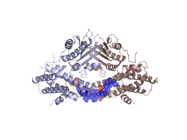
Structural Details of PDB entry 2BTZ
Structural Details of PDB entry 2BTZ
PDBid Chains Hinge Swapped Domain
2BTZ
A,B
A:362-372,B:362-372
A:373-384,B:373-384
Swapped-domain interface residues and interactions:
Chains Residues
A
362 , 363 , 364 , 365 , 367 , 369 , 377 , 378 , 380 , 381 , 382 , 383 , 384 ,
B
362 , 363 , 364 , 365 , 367 , 369 , 377 , 378 , 380 , 381 , 382 , 383 , 384 ,
Non-swapped-domain interface residues and interactions:
Chains Residues
A
22 , 149 , 152 , 219 , 221 , 268 , 270 , 272 , 273 , 274 , 275 , 277 , 279 , 288 , 289 , 290 , 335 , 337 , 339 , 340 , 341 , 342 , 343 , 344 , 347 , 351 , 353 , 372 ,
B
6 , 22 , 149 , 152 , 219 , 221 , 268 , 270 , 272 , 273 , 274 , 275 , 277 , 279 , 288 , 289 , 290 , 335 , 337 , 339 , 340 , 341 , 342 , 343 , 344 , 347 , 351 , 353 , 372 ,
Mutations in critical regions:
Chains
Hinge
Domain swapped interface Non-swapped interface Swapped Domain
A No mutation No mutation No mutation No mutation B No mutation No mutation No mutation No mutation
HIDE output:
JMOL Visualization:
2D-plot:
JOY Structural annotation for hinge hinge and swapped domain:
JOY output: