Pfam Domains mapped on to the structure: 2BS1
No.
Chain ID
Pfam ID
Pfam Description
Linkout - Pfam
Linkout - CDD
1
A
PF01819
Levivirus coat protein
PF01819
PF01819
Conserved Domain Database Superfamily Annotations: 2BS1
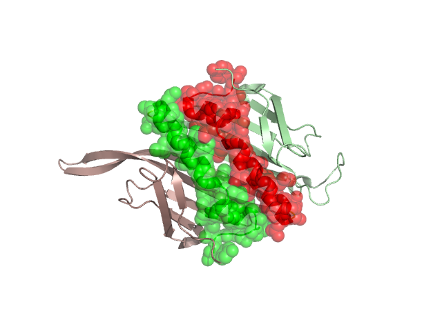
Structural Details of PDB entry 2BS1
Structural Details of PDB entry 2BS1
PDBid Chains Hinge Swapped Domain
2BS1
A,C,B
A:93-96,C:93-96,B:93-96
A:97-129,C:97-129,B:97-129
Swapped-domain interface residues and interactions:
Chains Residues
A
1 , 2 , 3 , 4 , 5 , 7 , 8 , 9 , 10 , 11 , 12 , 25 , 30 , 32 , 42 , 44 , 46 , 60 , 62 , 64 , 66 , 82 , 83 , 84 , 85 , 86 , 87 , 88 , 90 , 96 , 97 , 98 , 100 , 101 , 102 , 103 , 104 , 105 , 106 , 107 , 108 , 109 , 110 , 111 , 112 , 113 , 114 , 116 , 117 , 118 , 119 , 120 , 121 , 122 , 123 , 124 , 125 , 126 , 127 , 128 , 129 ,
B
1 , 2 , 3 , 4 , 7 , 8 , 9 , 10 , 11 , 12 , 25 , 30 , 32 , 42 , 44 , 46 , 48 , 56 , 58 , 60 , 62 , 64 , 66 , 67 , 68 , 84 , 85 , 86 , 87 , 88 , 89 , 90 , 91 , 93 , 95 , 96 , 98 , 100 , 101 , 102 , 103 , 104 , 105 , 106 , 107 , 108 , 109 , 110 , 111 , 112 , 113 , 114 , 116 , 117 , 118 , 119 , 120 , 121 , 122 , 123 , 124 , 125 , 126 , 127 , 128 ,
Non-swapped-domain interface residues and interactions:
Chains Residues
A
48 , 54 , 55 , 56 , 58 , 89 , 91 , 93 , 95 ,
B
78 , 80 , 81 , 83 ,
Mutations in critical regions:
Chains
Hinge
Domain swapped interface Non-swapped interface Swapped Domain
A No mutation ALA(87)ASN, LYS(89)GLU, No mutation C No mutation No mutation No mutation No mutation B No mutation ALA(87)ASN, LYS(89)GLU, No mutation No mutation
HIDE output:
JMOL Visualization:
2D-plot:
JOY Structural annotation for hinge hinge and swapped domain:
JOY output: