Home BrowseSearch Help Group

Pfam Domains mapped on to the structure: 2BNZ

No. Chain ID Pfam ID Pfam Description Linkout - Pfam Linkout - CDD
1 A PF07764 Omega Transcriptional Repressor PF07764 PF07764

Conserved Domain Database Superfamily Annotations: 2BNZ

No. PDB ID PSSM ID CDD Accession Superfamily Short Name Linkout - CDD
1 2BNZ 149046 Omega_Repress superfamily N -
Structural Details of PDB entry 2BNZ

Structural Details of PDB entry 2BNZ

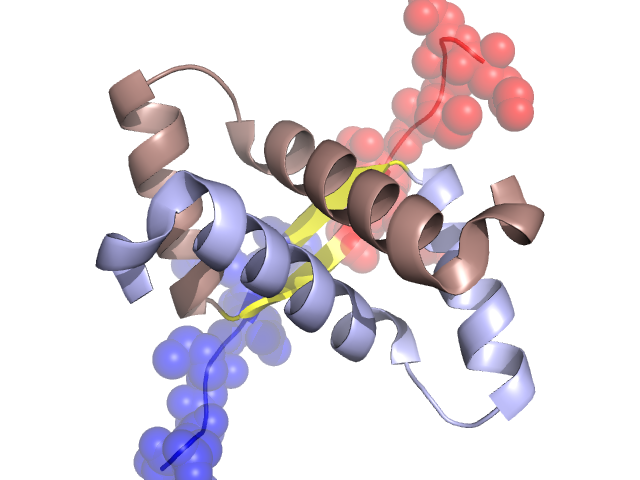
PDBid Chains Hinge Swapped Domain
2BNZ A,B A:30-33,B:30-33 A:22-29,B:22-29

Swapped-domain interface residues and interactions:

Chains Residues
A 25, 26, 27, 28, 29, 30, 31, 32, 33, 34, 37,
B 23, 25, 26, 27, 28, 29, 30, 31, 32, 33, 34, 37,

Non-swapped-domain interface residues and interactions:

Chains Residues
A 35, 36, 39, 40, 43, 46, 51, 52, 54, 55, 56, 57, 58, 59, 60, 61, 62, 63, 65, 66, 67, 70, 71,
B 35, 36, 39, 40, 41, 43, 52, 54, 55, 56, 57, 58, 59, 60, 61, 62, 63, 65, 66, 67, 70,

Swapped domains are represented using trasperent spheres.
Non-swapped part is represented using light color and cartoon representation.
Hinge region is shown in yellow color.

Mutations in critical regions:

Chains Hinge Domain swapped interface Non-swapped interface Swapped Domain
ANo mutationNo mutationNo mutationNo mutation
BNo mutationNo mutationNo mutationNo mutation

HIDE output:

Homologues found through HIDE algorithm

JMOL Visualization:

Jmol_logo            Jmol_logo            Jmol_logo

2D-plot:

A:2BNZ B:2BNZ

JOY Structural annotation for hinge hinge and swapped domain:

Hinge Swapped domain

JOY output:

ali file:2BNZ.ali
atm file:2BNZ.atm
cof file:2BNZ.cof
hbd file:2BNZ.hbd
html file:2BNZ.html
pdb file:2BNZ.pdb
ps file:2BNZ.ps
psa file:2BNZ.psa
rtf file:2BNZ.rtf
sst file:2BNZ.sst
tem file:2BNZ.tem