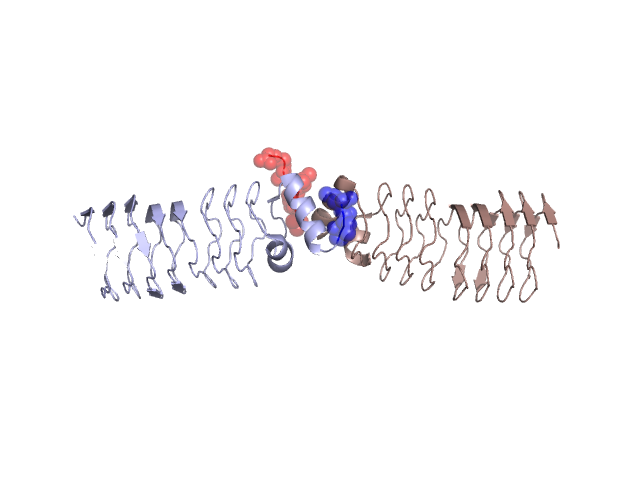
Pfam Domains mapped on to the structure: 2BM5 No. Chain ID Pfam ID Pfam Description Linkout - Pfam Linkout - CDD 1 A PF13599 Pentapeptide repeats (9 copies) PF13599 PF13599 2 A PF13599 Pentapeptide repeats (9 copies) PF13599 PF13599 3 A PF00805 Pentapeptide repeats (8 copies) PF00805 PF00805 4 A PF00805 Pentapeptide repeats (8 copies) PF00805 PF00805 Conserved Domain Database Superfamily Annotations: 2BM5 No. PDB ID PSSM ID CDD Accession Superfamily Short Name Linkout - CDD 1 2BM5 109845 cl02971 Pentapeptide superfamily - - 2 2BM5 109845 cl02971 Pentapeptide superfamily - - 3 2BM5 109845 cl02971 Pentapeptide superfamily - - Structural Details of PDB entry 2BM5 Structural Details of PDB entry 2BM5 PDBid Chains Hinge Swapped Domain 2BM5 A,B A:176-81,B:176-81 A:178-183,B:178-183 Swapped-domain interface residues and interactions: Chains Residues A 159, 160, 161, 162, 163, 164, 178, 179, 180, 181, 182, 183, B 159, 160, 161, 162, 163, 164, 165, 166, 169, 178, 179, 180, Non-swapped-domain interface residues and interactions: Chains Residues A 166, 169, 170, 172, 173, 176, 177, B 125, 145, 170, 172, 173, 176, 177, Swapped domains are represented using trasperent spheres. Non-swapped part is represented using light color and cartoon representation. Hinge region is shown in yellow color. Mutations in critical regions: Chains Hinge Domain swapped interface Non-swapped interface Swapped Domain ANo mutationNo mutationNo mutationNo mutation BNo mutationNo mutationNo mutationNo mutation HIDE output: Homologues found through HIDE algorithm JMOL Visualization: 2D-plot: A:2BM5 B:2BM5 JOY Structural annotation for hinge hinge and swapped domain: Hinge Swapped domain JOY output: ali file:2BM5.ali atm file:2BM5.atm cof file:2BM5.cof hbd file:2BM5.hbd html file:2BM5.html pdb file:2BM5.pdb ps file:2BM5.ps psa file:2BM5.psa rtf file:2BM5.rtf sst file:2BM5.sst tem file:2BM5.tem