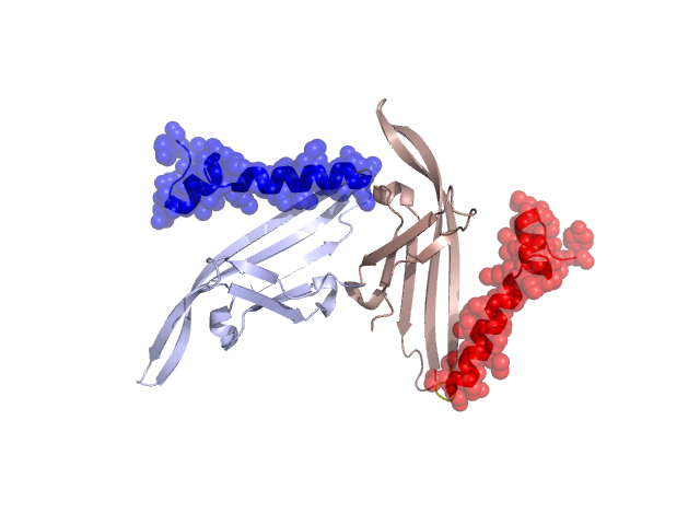
Pfam Domains mapped on to the structure: 2B2D No. Chain ID Pfam ID Pfam Description Linkout - Pfam Linkout - CDD 1 A PF01819 Levivirus coat protein PF01819 PF01819 Conserved Domain Database Superfamily Annotations: 2B2D No. PDB ID PSSM ID CDD Accession Superfamily Short Name Linkout - CDD 1 2B2D 133846 cl10174 cp superfamily - - Structural Details of PDB entry 2B2D Structural Details of PDB entry 2B2D PDBid Chains Hinge Swapped Domain 2B2D A,C,B A:93-96,C:93-96,B:93-96 A:97-129,C:97-129,B:97-129 Swapped-domain interface residues and interactions: Chains Residues A 35, 36, 37, 129, C 97, 98, Non-swapped-domain interface residues and interactions: Chains Residues A 2, 4, 5, 22, 24, 25, 26, 27, 38, 39, 77, 78, 79, C 1, 25, 27, 28, 48, 56, 94, 95, 96, Swapped domains are represented using trasperent spheres. Non-swapped part is represented using light color and cartoon representation. Hinge region is shown in yellow color. Mutations in critical regions: Chains Hinge Domain swapped interface Non-swapped interface Swapped Domain ANo mutationNo mutationNo mutationNo mutation CNo mutationNo mutationNo mutationNo mutation BNo mutationNo mutationNo mutationNo mutation HIDE output: Homologues found through HIDE algorithm JMOL Visualization: 2D-plot: A:2B2D B:2B2D C:2B2D JOY Structural annotation for hinge hinge and swapped domain: Hinge Swapped domain JOY output: ali file:2B2D.ali atm file:2B2D.atm cof file:2B2D.cof hbd file:2B2D.hbd html file:2B2D.html pdb file:2B2D.pdb ps file:2B2D.ps psa file:2B2D.psa rtf file:2B2D.rtf sst file:2B2D.sst tem file:2B2D.tem