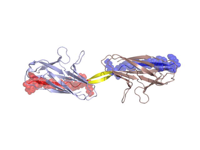
Structural Details of PDB entry 2AXW
Structural Details of PDB entry 2AXW
PDBid Chains Hinge Swapped Domain
2AXW
A,B
A:117-121,B:117-121
A:122-134,B:122-134
Swapped-domain interface residues and interactions:
Chains Residues
A
1 , 3 , 7 , 10 , 11 , 12 , 105 , 106 , 107 , 108 , 109 , 110 , 111 , 112 , 113 , 114 , 115 , 116 , 117 , 118 , 122 , 123 , 124 , 125 , 126 , 127 , 128 , 129 , 130 , 131 , 132 , 133 , 134 ,
B
1 , 3 , 7 , 105 , 106 , 107 , 108 , 109 , 110 , 111 , 112 , 113 , 114 , 115 , 116 , 117 , 122 , 123 , 124 , 125 , 126 , 127 , 128 , 129 , 130 , 131 , 132 , 133 ,
Non-swapped-domain interface residues and interactions:
Chains Residues
A
5 , 21 , 22 , 30 , 33 , 36 , 38 , 40 , 103 , 119 , 120 , 121 ,
B
5 , 12 , 13 , 15 , 21 , 30 , 33 , 36 , 38 , 40 , 103 , 118 , 119 , 120 , 121 ,