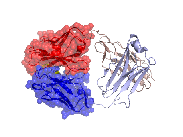
Structural Details of PDB entry 2AGJ
Structural Details of PDB entry 2AGJ
PDBid Chains Hinge Swapped Domain
2AGJ
H,L
H:105-109,L:95-98
H:2-104,L:1-94
Swapped-domain interface residues and interactions:
Chains Residues
H
41 , 45 , 46 , 47 , 49 , 52 , 54 , 60 , 61 , 63 , 96 , 103 , 104 , 105 , 106 , 107 , 108 , 110 , 112 ,
L
33 , 37 , 39 , 42 , 43 , 44 , 45 , 47 , 50 , 56 , 57 , 88 , 92 , 95 , 96 , 97 , 99 , 101 ,
Non-swapped-domain interface residues and interactions:
Chains Residues
H
129 , 130 , 131 , 132 , 133 , 134 , 135 , 142 , 144 , 148 , 150 , 172 , 173 , 174 , 175 , 176 , 178 , 180 , 187 , 189 , 191 , 218 ,
L
117 , 118 , 119 , 120 , 122 , 124 , 125 , 128 , 132 , 134 , 136 , 138 , 139 , 161 , 163 , 164 , 165 , 168 , 175 , 177 , 179 , 181 , 208 , 215 ,