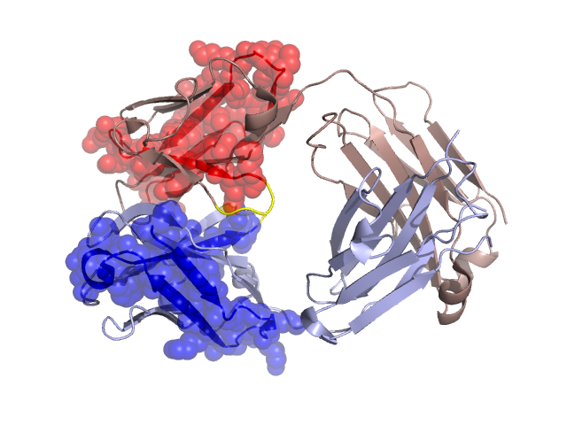
Structural Details of PDB entry 2ADI
Structural Details of PDB entry 2ADI
PDBid Chains Hinge Swapped Domain
2ADI
A,B
A:40-43,B:41-44
A:1-39,B:1-40
Swapped-domain interface residues and interactions:
Chains Residues
A
36 , 38 ,
B
39 ,
Non-swapped-domain interface residues and interactions:
Chains Residues
A
43 , 44 , 46 , 49 , 50 , 55 , 87 , 89 , 91 , 94 , 95 , 96 , 98 , 116 , 118 , 119 , 120 , 121 , 122 , 123 , 124 , 127 , 131 , 133 , 135 , 137 , 138 , 160 , 161 , 162 , 163 , 164 , 167 , 174 , 176 , 178 , 180 , 211 ,
B
44 , 45 , 47 , 59 , 61 , 91 , 100 , 101 , 103 , 104 , 105 , 122 , 123 , 124 , 125 , 126 , 137 , 141 , 143 , 164 , 165 , 166 , 167 , 169 , 171 , 176 , 178 , 180 , 182 , 208 ,