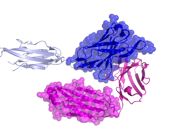
Pfam Domains mapped on to the structure: 1ZLS No. Chain ID Pfam ID Pfam Description Linkout - Pfam Linkout - CDD 1 L PF13927 Immunoglobulin domain PF13927 PF13927 2 L PF13927 Immunoglobulin domain PF13927 PF13927 Conserved Domain Database Superfamily Annotations: 1ZLS No. PDB ID PSSM ID CDD Accession Superfamily Short Name Linkout - CDD 1 1ZLS 212623 cl11960 Ig superfamily - - 2 1ZLS 143186 cd04985 IgC_CH1 - cl11960 3 1ZLS 212623 cl11960 Ig superfamily - - 4 1ZLS 212623 cl11960 Ig superfamily - - Structural Details of PDB entry 1ZLS Structural Details of PDB entry 1ZLS PDBid Chains Hinge Swapped Domain 1ZLS H,A,L,B H:113-113,A:113-113,L:113-113,B:113-113 H:1-112,A:1-112,L:114-210,B:1-112 Swapped-domain interface residues and interactions: Chains Residues H 33, 39, 43, 44, 45, 47, 50, 52, 56, 58, 89, 91, 95, 97, 98, 100, 101, 103, 104, 105, L 43, 44, 46, 94, 95, 96, 98, 100, 210, Non-swapped-domain interface residues and interactions: Chains Residues L 32, 36, 38, 40, 41, 49, 56, 85, 87, 89, 91, 92, 93, 99, Swapped domains are represented using trasperent spheres. Non-swapped part is represented using light color and cartoon representation. Hinge region is shown in yellow color. Mutations in critical regions: Chains Hinge Domain swapped interface Non-swapped interface Swapped Domain HNo mutationNo mutationNo mutationNo mutation ANo mutationNo mutationNo mutationNo mutation LNo mutationNo mutationNo mutationNo mutation BNo mutationNo mutationNo mutationNo mutation HIDE output: Homologues found through HIDE algorithm JMOL Visualization: 2D-plot: A:1ZLS B:1ZLS H:1ZLS L:1ZLS JOY Structural annotation for hinge hinge and swapped domain: Hinge Swapped domain JOY output: ali file:1ZLS.ali atm file:1ZLS.atm cof file:1ZLS.cof hbd file:1ZLS.hbd html file:1ZLS.html pdb file:1ZLS.pdb ps file:1ZLS.ps psa file:1ZLS.psa rtf file:1ZLS.rtf sst file:1ZLS.sst tem file:1ZLS.tem