Pfam Domains mapped on to the structure: 1Z7W
No.
Chain ID
Pfam ID
Pfam Description
Linkout - Pfam
Linkout - CDD
1
A
PF00291
Pyridoxal-phosphate dependent enzyme
PF00291
PF00291
Conserved Domain Database Superfamily Annotations: 1Z7W
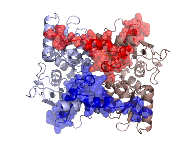
Structural Details of PDB entry 1Z7W
Structural Details of PDB entry 1Z7W
PDBid Chains Hinge Swapped Domain
1Z7W
A,B
A:263-266,B:263-266
A:267-322,B:267-322
Swapped-domain interface residues and interactions:
Chains Residues
A
99 , 103 , 106 , 107 , 110 , 111 , 117 , 118 , 119 , 120 , 298 , 300 , 301 , 303 , 308 , 311 , 312 , 314 , 315 , 316 , 318 , 319 , 320 , 321 , 322 ,
B
99 , 103 , 106 , 107 , 110 , 111 , 117 , 118 , 119 , 120 , 298 , 300 , 301 , 303 , 308 , 311 , 312 , 314 , 315 , 316 , 318 , 319 , 320 , 321 ,
Non-swapped-domain interface residues and interactions:
Chains Residues
A
3 , 4 , 5 , 6 , 7 , 8 , 9 , 12 , 17 , 18 , 19 , 20 , 22 , 33 , 36 , 39 , 41 , 42 , 43 , 87 , 88 , 104 , 108 , 112 , 113 , 116 , 121 , 123 , 131 , 167 , 168 , 169 , 259 , 260 , 261 , 262 , 263 , 264 , 265 ,
B
3 , 4 , 5 , 6 , 7 , 8 , 9 , 12 , 17 , 18 , 19 , 20 , 22 , 33 , 36 , 39 , 41 , 42 , 43 , 87 , 88 , 104 , 108 , 112 , 113 , 116 , 121 , 123 , 131 , 167 , 168 , 169 , 259 , 260 , 261 , 262 , 263 , 264 , 265 ,
Mutations in critical regions:
Chains
Hinge
Domain swapped interface Non-swapped interface Swapped Domain
A No mutation No mutation No mutation No mutation B No mutation No mutation No mutation No mutation
HIDE output:
JMOL Visualization:
2D-plot:
JOY Structural annotation for hinge hinge and swapped domain:
JOY output: