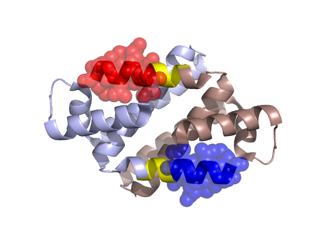
Structural Details of PDB entry 1YVW
Structural Details of PDB entry 1YVW
PDBid Chains Hinge Swapped Domain
1YVW
A,B
A:83-86,B:83-86
A:87-95,B:87-95
Swapped-domain interface residues and interactions:
Chains Residues
A
4 , 8 , 61 , 64 , 87 , 88 , 89 , 91 , 92 , 95 ,
B
4 , 8 , 61 , 64 , 87 , 88 , 89 , 91 , 92 ,
Non-swapped-domain interface residues and interactions:
Chains Residues
A
5 , 7 , 11 , 15 , 25 , 33 , 34 , 37 , 40 , 41 , 44 , 47 , 48 , 51 , 52 , 54 , 56 , 59 , 60 , 65 , 66 , 67 , 68 , 70 , 71 , 73 , 74 , 77 , 78 , 79 , 83 , 84 ,
B
5 , 7 , 11 , 15 , 25 , 33 , 34 , 37 , 40 , 41 , 44 , 47 , 48 , 51 , 52 , 54 , 56 , 59 , 60 , 65 , 66 , 67 , 68 , 70 , 71 , 73 , 74 , 77 , 78 , 79 , 83 , 84 ,