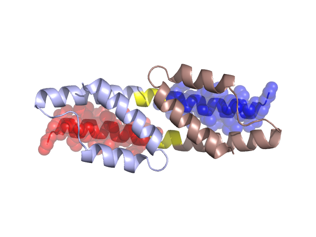
Structural Details of PDB entry 1YBZ
Structural Details of PDB entry 1YBZ
PDBid Chains Hinge Swapped Domain
1YBZ
A,B
A:18-22,B:18-22
A:0-17,B:0-17
Swapped-domain interface residues and interactions:
Chains Residues
A
1 , 3 , 4 , 7 , 8 , 10 , 11 , 12 , 14 , 15 , 16 , 17 , 21 , 25 , 28 , 32 ,
B
1 , 3 , 4 , 7 , 8 , 10 , 11 , 12 , 14 , 15 , 16 , 17 , 21 , 25 , 28 , 32 ,
Non-swapped-domain interface residues and interactions:
Chains Residues
A
18 , 19 , 22 , 23 , 24 , 26 , 31 , 35 , 36 , 39 , 41 , 45 , 48 , 55 , 56 , 58 , 59 , 61 , 62 , 63 , 65 , 66 , 69 , 75 ,
B
18 , 19 , 22 , 23 , 24 , 26 , 31 , 35 , 36 , 39 , 41 , 45 , 48 , 55 , 56 , 58 , 59 , 61 , 62 , 63 , 65 , 66 , 69 ,