Pfam Domains mapped on to the structure: 1YB4
No.
Chain ID
Pfam ID
Pfam Description
Linkout - Pfam
Linkout - CDD
1
A
PF03446
NAD binding domain of 6-phosphogluconate dehydrogenase
PF03446
PF03446
2
A
PF03807
NADP oxidoreductase coenzyme F420-dependent
PF03807
PF03807
Conserved Domain Database Superfamily Annotations: 1YB4
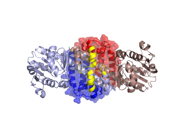
Structural Details of PDB entry 1YB4
Structural Details of PDB entry 1YB4
PDBid Chains Hinge Swapped Domain
1YB4
A,B
A:206-220,B:206-220
A:221-291,B:221-291
Swapped-domain interface residues and interactions:
Chains Residues
A
185 , 189 , 245 , 249 , 250 , 251 , 252 , 253 , 254 , 255 , 258 , 262 , 283 , 287 , 291 ,
B
185 , 189 , 192 , 245 , 249 , 250 , 251 , 252 , 253 , 254 , 255 , 258 , 262 , 283 , 287 ,
Non-swapped-domain interface residues and interactions:
Chains Residues
A
97 , 101 , 132 , 155 , 157 , 158 , 159 , 160 , 164 , 167 , 168 , 170 , 171 , 172 , 174 , 175 , 176 , 178 , 179 , 181 , 182 , 183 , 186 , 190 , 192 , 193 , 194 , 195 , 199 , 200 , 203 , 204 , 206 , 207 , 208 , 209 , 210 , 211 , 212 , 213 , 216 , 217 ,
B
97 , 101 , 132 , 155 , 157 , 158 , 159 , 160 , 164 , 167 , 168 , 170 , 171 , 172 , 174 , 175 , 176 , 178 , 179 , 181 , 182 , 183 , 186 , 190 , 193 , 194 , 195 , 199 , 200 , 203 , 204 , 206 , 207 , 208 , 209 , 210 , 211 , 212 , 213 , 216 , 217 ,
Mutations in critical regions:
Chains
Hinge
Domain swapped interface Non-swapped interface Swapped Domain
A No mutation No mutation No mutation No mutation B No mutation No mutation No mutation No mutation
HIDE output:
JMOL Visualization:
2D-plot:
JOY Structural annotation for hinge hinge and swapped domain:
JOY output: