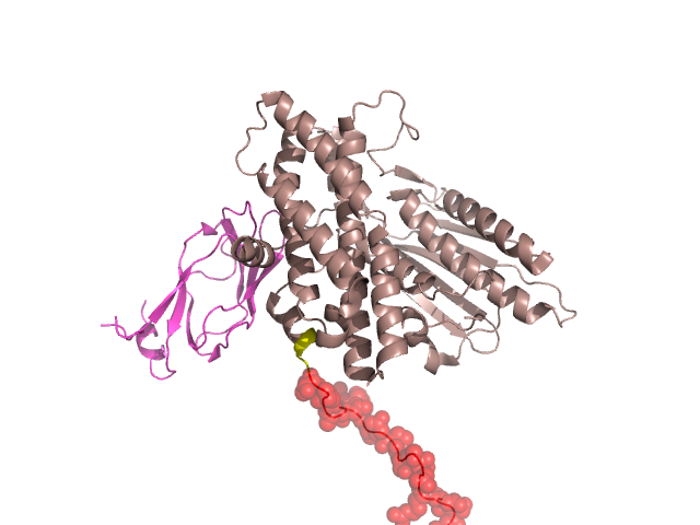
Pfam Domains mapped on to the structure: 1Y8P No. Chain ID Pfam ID Pfam Description Linkout - Pfam Linkout - CDD 1 A PF10436 Mitochondrial branched-chain alpha-ketoacid dehydrogenase kinase PF10436 PF10436 2 A PF02518 Histidine kinase-, DNA gyrase B-, and HSP90-like ATPase PF02518 PF02518 3 B PF00364 Biotin-requiring enzyme PF00364 PF00364 Conserved Domain Database Superfamily Annotations: 1Y8P No. PDB ID PSSM ID CDD Accession Superfamily Short Name Linkout - CDD 1 1Y8P 133458 cd06849 lipoyl_domain - cl11404 2 1Y8P 209306 cl11404 Biotinyl_lipoyl_domains superfamily - - Structural Details of PDB entry 1Y8P Structural Details of PDB entry 1Y8P PDBid Chains Hinge Swapped Domain 1Y8P A,D,C,B A:379-382,D:379-382,C:379-382,B:379-382 A:383-406,D:383-406,C:383-406,B:383-406 Swapped-domain interface residues and interactions: Chains Residues A 401, Non-swapped-domain interface residues and interactions: Chains Residues A 22, 23, 25, 26, 27, 31, 48, 51, 55, 373, 374, 378, B 139, 140, 141, 142, 143, 162, 163, 164, 173, 174, 176, 179, Swapped domains are represented using trasperent spheres. Non-swapped part is represented using light color and cartoon representation. Hinge region is shown in yellow color. Mutations in critical regions: Chains Hinge Domain swapped interface Non-swapped interface Swapped Domain ANo mutationNo mutationNo mutationNo mutation DNo mutationNo mutationNo mutationNo mutation CNo mutationNo mutationNo mutationNo mutation BNo mutationNo mutationNo mutationNo mutation HIDE output: Homologues found through HIDE algorithm JMOL Visualization: 2D-plot: D:1Y8P A:1Y8P B:1Y8P C:1Y8P JOY Structural annotation for hinge hinge and swapped domain: Hinge Swapped domain JOY output: ali file:1Y8P.ali atm file:1Y8P.atm cof file:1Y8P.cof hbd file:1Y8P.hbd html file:1Y8P.html pdb file:1Y8P.pdb ps file:1Y8P.ps psa file:1Y8P.psa rtf file:1Y8P.rtf sst file:1Y8P.sst tem file:1Y8P.tem