Pfam Domains mapped on to the structure: 1Y3H
No.
Chain ID
Pfam ID
Pfam Description
Linkout - Pfam
Linkout - CDD
1
A
PF01513
ATP-NAD kinase
PF01513
PF01513
Conserved Domain Database Superfamily Annotations: 1Y3H
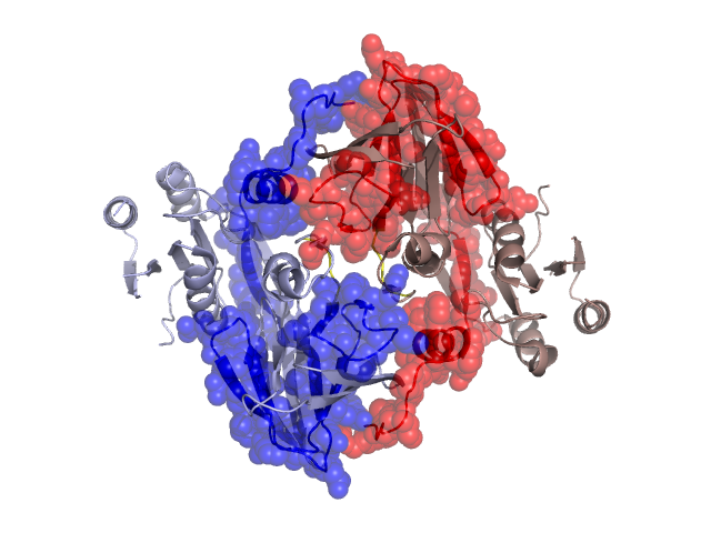
Structural Details of PDB entry 1Y3H
Structural Details of PDB entry 1Y3H
PDBid Chains Hinge Swapped Domain
1Y3H
A,B
A:214-218,B:214-218
A:219-307,B:219-307
Swapped-domain interface residues and interactions:
Chains Residues
A
175 , 181 , 182 , 183 , 184 , 185 , 209 , 210 , 211 , 212 , 214 , 215 , 229 , 231 , 232 , 233 , 234 , 235 , 236 , 244 , 247 , 267 , 269 , 288 , 290 , 291 , 293 , 294 , 299 , 300 , 301 , 302 , 303 , 304 , 305 , 306 , 307 ,
B
175 , 178 , 180 , 181 , 182 , 183 , 184 , 185 , 209 , 210 , 211 , 212 , 214 , 215 , 227 , 229 , 231 , 232 , 233 , 234 , 235 , 236 , 244 , 247 , 267 , 269 , 271 , 288 , 290 , 291 , 293 , 294 , 299 , 300 , 301 , 302 , 303 , 304 , 305 , 306 ,
Non-swapped-domain interface residues and interactions:
Chains Residues
A
146 , 149 , 177 , 178 , 179 , 180 , 204 , 207 , 208 , 217 ,
B
146 , 177 , 204 , 207 , 208 , 217 ,
Mutations in critical regions:
Chains
Hinge
Domain swapped interface Non-swapped interface Swapped Domain
A No mutation ILE(302)THR, No mutation ILE(302)THR, B No mutation ILE(302)THR, No mutation ILE(302)THR,
HIDE output:
JMOL Visualization:
2D-plot:
JOY Structural annotation for hinge hinge and swapped domain:
JOY output: