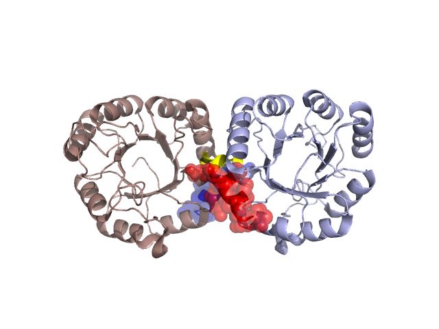
Structural Details of PDB entry 1Y0E
Structural Details of PDB entry 1Y0E
PDBid Chains Hinge Swapped Domain
1Y0E
A,B
A:207-210,B:207-210
A:211-221,B:211-221
Swapped-domain interface residues and interactions:
Chains Residues
A
6 , 31 , 34 , 35 , 186 , 189 , 190 , 206 , 212 , 213 , 214 , 215 , 216 , 217 , 219 , 221 ,
B
6 , 31 , 34 , 35 , 186 , 189 , 190 , 206 , 212 , 213 , 214 , 215 , 216 , 217 , 219 ,
Non-swapped-domain interface residues and interactions:
Chains Residues
A
17 , 18 , 20 , 22 , 23 , 26 , 29 , 30 , 33 , 202 , 205 , 209 , 210 ,
B
17 , 18 , 22 , 23 , 26 , 29 , 30 , 33 , 202 , 205 , 207 , 209 , 210 ,