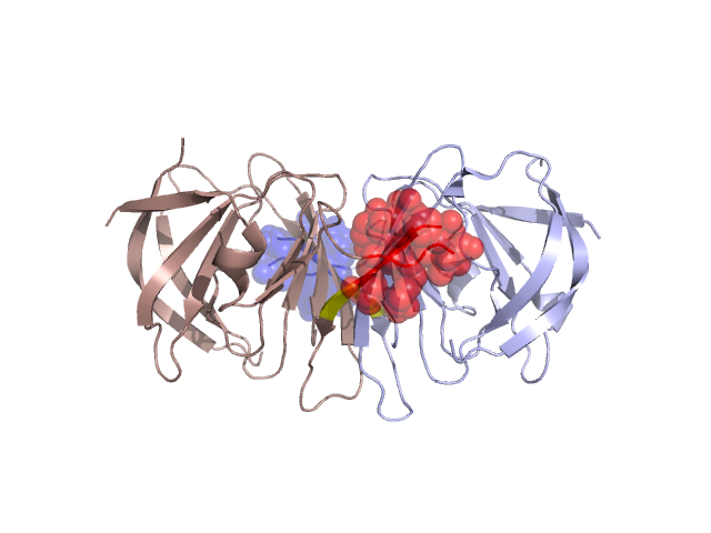
Structural Details of PDB entry 1XSQ
Structural Details of PDB entry 1XSQ
PDBid Chains Hinge Swapped Domain
1XSQ
A,B
A:22-26,B:22-26;A:40-42,B:40-42
A:27-39,B:27-39
Swapped-domain interface residues and interactions:
Chains Residues
A
35 , 36 , 37 , 38 , 55 , 56 , 57 , 58 , 59 , 60 ,
B
35 , 36 , 37 , 38 , 55 , 56 , 57 , 58 , 59 , 60 ,
Non-swapped-domain interface residues and interactions:
Chains Residues
A
20 , 22 , 40 , 41 , 42 , 43 , 44 , 50 , 51 , 52 , 53 , 54 , 82 , 85 , 86 , 113 , 127 , 137 , 139 , 142 , 160 ,
B
20 , 22 , 40 , 41 , 42 , 43 , 44 , 45 , 51 , 52 , 53 , 54 , 82 , 85 , 86 , 113 , 127 , 137 , 139 , 142 ,