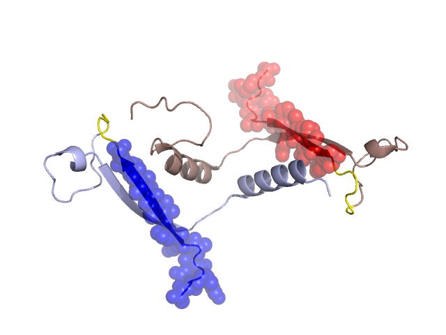
Pfam Domains mapped on to the structure: 1XG0 No. Chain ID Pfam ID Pfam Description Linkout - Pfam Linkout - CDD 1 A PF02972 Phycoerythrin, alpha/beta chain PF02972 PF02972 2 B PF02972 Phycoerythrin, alpha/beta chain PF02972 PF02972 3 C PF00502 Phycobilisome protein PF00502 PF00502 Gene Ontology Annotations: 1XG0 No. GO ID GO Description Linkout - AmiGO 1 GO:0030089 phycobilisome GO:0030089 Conserved Domain Database Superfamily Annotations: 1XG0 No. PDB ID PSSM ID CDD Accession Superfamily Short Name Linkout - CDD 1 1XG0 208826 cl08227 Phycobilisome superfamily - - Structural Details of PDB entry 1XG0 Structural Details of PDB entry 1XG0 PDBid Chains Hinge Swapped Domain 1XG0 A,B A:16-21,B:16-21 A:1-15,B:1-15 Swapped-domain interface residues and interactions: Chains Residues A 10, 12, 13, 14, 15, 59, 63, B 10, 11, 12, 13, 14, 15, 59, 62, 63, 66, Non-swapped-domain interface residues and interactions: Chains Residues A 16, 17, 18, 19, 50, 53, 56, 62, 64, 76, B 16, 17, 18, 19, 47, 48, 49, 50, 53, 65, Swapped domains are represented using trasperent spheres. Non-swapped part is represented using light color and cartoon representation. Hinge region is shown in yellow color. Mutations in critical regions: Chains Hinge Domain swapped interface Non-swapped interface Swapped Domain ANo mutationNo mutationNo mutationLYZ(4)LYS, BNo mutationNo mutationNo mutationNo mutation HIDE output: Homologues found through HIDE algorithm JMOL Visualization: 2D-plot: A:1XG0 B:1XG0 JOY Structural annotation for hinge hinge and swapped domain: Hinge Swapped domain JOY output: ali file:1XG0.ali atm file:1XG0.atm cof file:1XG0.cof hbd file:1XG0.hbd html file:1XG0.html pdb file:1XG0.pdb ps file:1XG0.ps psa file:1XG0.psa rtf file:1XG0.rtf sst file:1XG0.sst tem file:1XG0.tem