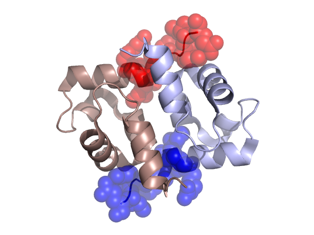
Pfam Domains mapped on to the structure: 1X2I No. Chain ID Pfam ID Pfam Description Linkout - Pfam Linkout - CDD 1 A PF12826 Helix-hairpin-helix motif PF12826 PF12826 2 A PF00633 Helix-hairpin-helix motif PF00633 PF00633 Gene Ontology Annotations: 1X2I No. GO ID GO Description Linkout - AmiGO 1 GO:0003677 DNA binding GO:0003677 Conserved Domain Database Superfamily Annotations: 1X2I No. PDB ID PSSM ID CDD Accession Superfamily Short Name Linkout - CDD 1 1X2I 209897 cl14786 ENDO3c superfamily - - 2 1X2I 209897 cl14786 ENDO3c superfamily - - Structural Details of PDB entry 1X2I Structural Details of PDB entry 1X2I PDBid Chains Hinge Swapped Domain 1X2I A,B A:42-27,B:42-27 A:61-69,B:61-69 Swapped-domain interface residues and interactions: Chains Residues A 5, 6, 28, 29, 32, 33, 34, 35, 61, 62, 63, 64, 65, 66, 67, 68, 69, B 5, 6, 9, 28, 29, 32, 33, 34, 35, 61, 62, 63, 64, 65, 66, 67, 68, Non-swapped-domain interface residues and interactions: Chains Residues A 8, 9, 10, 12, 15, 17, 25, 36, 38, 39, 60, B 8, 10, 11, 12, 15, 16, 17, 25, 38, 39, 60, Swapped domains are represented using trasperent spheres. Non-swapped part is represented using light color and cartoon representation. Hinge region is shown in yellow color. Mutations in critical regions: Chains Hinge Domain swapped interface Non-swapped interface Swapped Domain ANo mutationNo mutationNo mutationNo mutation BNo mutationNo mutationNo mutationNo mutation HIDE output: Homologues found through HIDE algorithm JMOL Visualization: 2D-plot: A:1X2I B:1X2I JOY Structural annotation for hinge hinge and swapped domain: Hinge Swapped domain JOY output: ali file:1X2I.ali atm file:1X2I.atm cof file:1X2I.cof hbd file:1X2I.hbd html file:1X2I.html pdb file:1X2I.pdb ps file:1X2I.ps psa file:1X2I.psa rtf file:1X2I.rtf sst file:1X2I.sst tem file:1X2I.tem