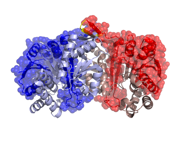
Structural Details of PDB entry 1X0L
Structural Details of PDB entry 1X0L
PDBid Chains Hinge Swapped Domain
1X0L
A,B
A:133-136,B:133-136
A:2-132,B:2-132
Swapped-domain interface residues and interactions:
Non-swapped-domain interface residues and interactions:
Chains Residues
A
134 , 135 , 136 , 137 , 138 , 139 , 140 , 141 , 142 , 143 , 144 , 171 , 174 , 175 , 176 , 178 , 181 , 203 , 204 , 207 , 208 , 211 , 212 , 225 , 226 , 228 , 229 , 232 , 233 , 236 , 237 , 239 , 240 , 241 , 334 ,
B
134 , 135 , 136 , 137 , 138 , 139 , 140 , 141 , 142 , 143 , 144 , 171 , 175 , 178 , 181 , 203 , 204 , 205 , 207 , 208 , 211 , 212 , 225 , 228 , 229 , 232 , 233 , 236 , 237 , 240 , 241 ,