Home BrowseSearch Help Group

Pfam Domains mapped on to the structure: 1VQ3

No. Chain ID Pfam ID Pfam Description Linkout - Pfam Linkout - CDD
1 A PF02700 Phosphoribosylformylglycinamidine (FGAM) synthase PF02700 PF02700

Conserved Domain Database Superfamily Annotations: 1VQ3

No. PDB ID PSSM ID CDD Accession Superfamily Short Name Linkout - CDD
1 1VQ3 207199 cl00789 PurS superfamily - -
Structural Details of PDB entry 1VQ3

Structural Details of PDB entry 1VQ3

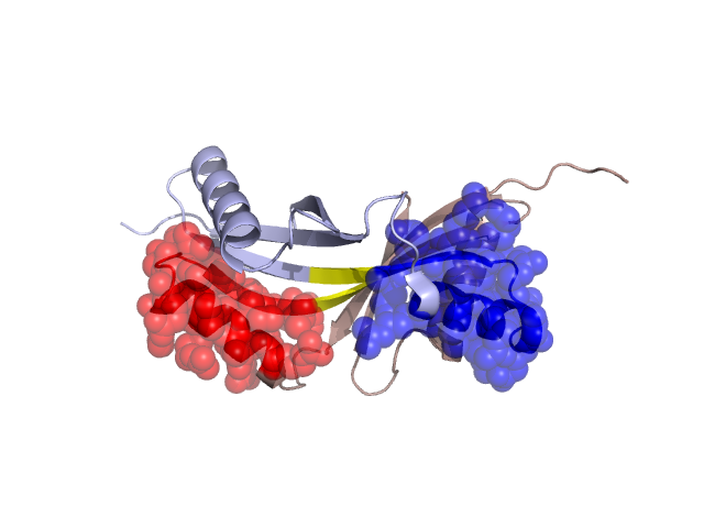
PDBid Chains Hinge Swapped Domain
1VQ3 A,B A:12-19,B:12-19;A:42-44,B:42-44 A:20-41,B:20-41

Swapped-domain interface residues and interactions:

Chains Residues
A 24, 27, 28, 32, 34, 35, 36, 37, 38, 39, 40, 41, 44, 45, 46, 47, 48, 49, 50, 60, 64,
B 24, 27, 28, 32, 34, 35, 36, 37, 38, 39, 40, 41, 44, 45, 46, 47, 48, 49, 50, 60, 64,

Non-swapped-domain interface residues and interactions:

Chains Residues
A 3, 9, 11, 42, 43, 63, 67, 68, 69, 78, 80, 82,
B 3, 9, 11, 42, 43, 63, 67, 68, 69, 78, 80,

Swapped domains are represented using trasperent spheres.
Non-swapped part is represented using light color and cartoon representation.
Hinge region is shown in yellow color.

Mutations in critical regions:

Chains Hinge Domain swapped interface Non-swapped interface Swapped Domain
ANo mutationNo mutationNo mutationNo mutation
BNo mutationNo mutationNo mutationNo mutation

HIDE output:

Homologues found through HIDE algorithm

JMOL Visualization:

Jmol_logo            Jmol_logo            Jmol_logo

2D-plot:

A:1VQ3 B:1VQ3

JOY Structural annotation for hinge hinge and swapped domain:

Hinge Swapped domain

JOY output:

ali file:1VQ3.ali
atm file:1VQ3.atm
cof file:1VQ3.cof
hbd file:1VQ3.hbd
html file:1VQ3.html
pdb file:1VQ3.pdb
ps file:1VQ3.ps
psa file:1VQ3.psa
rtf file:1VQ3.rtf
sst file:1VQ3.sst
tem file:1VQ3.tem