Home BrowseSearch Help Group

Pfam Domains mapped on to the structure: 1VPW

No. Chain ID Pfam ID Pfam Description Linkout - Pfam Linkout - CDD
1 A PF00356 Bacterial regulatory proteins, lacI family PF00356 PF00356
2 A PF00532 Periplasmic binding proteins and sugar binding domain of LacI family PF00532 PF00532
3 A PF13407 Periplasmic binding protein domain PF13407 PF13407
4 A PF13377 Periplasmic binding protein-like domain PF13377 PF13377

Gene Ontology Annotations: 1VPW

No. GO ID GO Description Linkout - AmiGO
1 GO:0005622 intracellular GO:0005622

Conserved Domain Database Superfamily Annotations: 1VPW

No. PDB ID PSSM ID CDD Accession Superfamily Short Name Linkout - CDD
1 1VPW 107270 cd06275 PBP1_PurR - cl10011
2 1VPW 209116 cl10011 Periplasmic_Binding_Protein_Type_1 superfamily - -
3 1VPW 143331 cd01392 HTH_LacI - cl15761
4 1VPW 210161 cl15761 HTH_XRE superfamily - -
5 1VPW 210161 cl15761 HTH_XRE superfamily - -
Structural Details of PDB entry 1VPW

Structural Details of PDB entry 1VPW

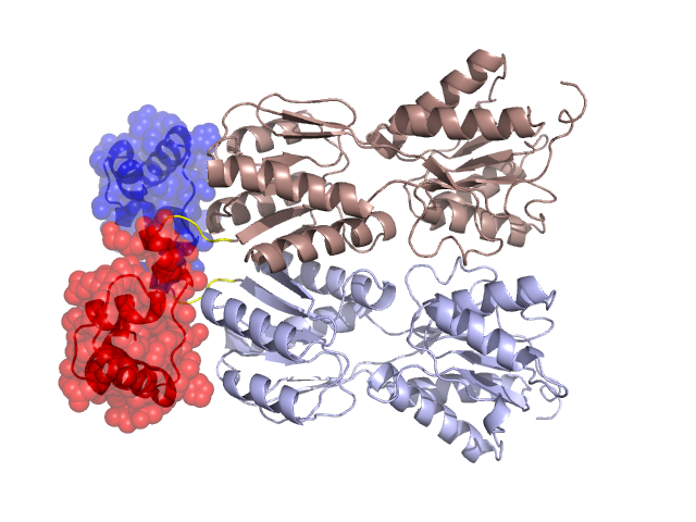
PDBid Chains Hinge Swapped Domain
1VPW A,B A:57-60,B:57-60 A:3-56,B:3-56

Swapped-domain interface residues and interactions:

Chains Residues
A 3, 44, 46, 47, 48, 49, 50, 51, 53, 54, 112, 113, 114,
B 3, 44, 46, 47, 48, 49, 50, 51, 53, 54, 112, 113, 114,

Non-swapped-domain interface residues and interactions:

Chains Residues
A 59, 69, 70, 75, 76, 78, 79, 82, 83, 86, 89, 91, 92, 93, 94, 96, 98, 103, 106, 107, 110, 115, 136, 222, 223, 224, 227, 249, 252, 253, 256, 259, 260, 264, 278, 279, 281, 282, 284, 285, 329, 340,
B 59, 69, 70, 75, 76, 78, 79, 82, 83, 86, 89, 91, 92, 93, 94, 96, 98, 103, 106, 107, 110, 115, 136, 222, 223, 224, 227, 249, 252, 253, 256, 259, 260, 264, 278, 279, 281, 282, 284, 285, 329,

Swapped domains are represented using trasperent spheres.
Non-swapped part is reprented using light color and cartoon reprentation.
Hinge region is shown in yellow color.

Mutations in critical regions:

Chains Hinge Domain swapped interface Non-swapped interface Swapped Domain
ANo mutationNo mutationNo mutationNo mutation
BNo mutationNo mutationNo mutationNo mutation

HIDE output:

Homologues found through HIDE algorithm

JMOL Visualization:

Jmol_logo Jmol_logo

2D-plot:

3:1VPW A:1VPW B:1VPW

JOY Structural annotation for hinge hinge and swapped domain:

Hinge Swapped domain

JOY output:

ali file:1VPW.ali
atm file:1VPW.atm
cof file:1VPW.cof
hbd file:1VPW.hbd
html file:1VPW.html
pdb file:1VPW.pdb
ps file:1VPW.ps
psa file:1VPW.psa
rtf file:1VPW.rtf
sst file:1VPW.sst
tem file:1VPW.tem